This post is an answer to the Case – 42-year-old Woman With Shortness of Breath
Findings
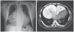
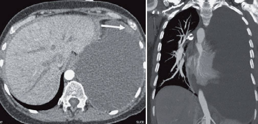
- Two-view chest radiograph (images below) shows increased opacification of the lower and lateral left hemothorax with a meniscus.
- The mediastinum is shift ed rightward and the diaphragm is shifted inferiorly. The mass effect is better seen on the subsequent CT (images below). A small nodule is seen along the anterior pleura (arrow in first image).
Differential Diagnosis
Unilateral pleural effusions are usually seen in infection (empyema), malignancy, and trauma (hemothorax). An abdominal process may present with a large unilateral effusion, such as a large right effusion in the setting of cirrhosis (hepatic hydrothorax). Other less common causes of large unilateral effusions include chylothorax, and glucothorax (from an extravascular placement of a central venous catheter).
Teaching Points
- Large unilateral effusions are rare in the setting of congestive heart failure and should prompt consideration of other potential etiologies.
- A large effusion may exert tension on surrounding structures and may have the same physiologic significance as a tension pneumothorax.
- A decubitus radiograph (side of effusion down) will show that the effusion is mobile when the effusion is small or medium. In larger effusions, there may be little appreciable change on a decubitus film.
Management
- Effusions under tension require drainage on an urgent/emergent basis.
- If the patient is hemodynamically stable, a CT may be performed for further characterization and thoracostomy tube planning.
SIMILAR CASES:
Further Reading
Johnson JL. Pleural effusions in cardiovascular disease. Pearls for correlating the evidence with the cause. Postgrad Med. 2000 Apr; 107 (4): 95-101 .