This post is an answer to the ECG Case 306
Key features
- Regular narrow complex tachycardia
- Rate ~ 130 bpm
- Left axis deviation
- Late R wave transition
- Relatively flat isoelectric line
- Possible atrial activity seen in lead III and V6
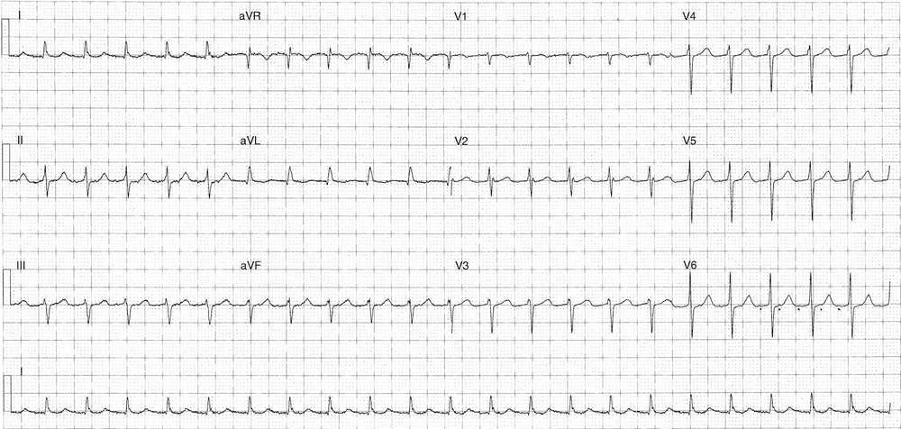
Differential diagnosis
- Atrial flutter
- Atrial tachycardia
- Accelerated junctional rhythm
This following ECG was taken during treatment with adenosine, hence the dramatic ventricular pause.
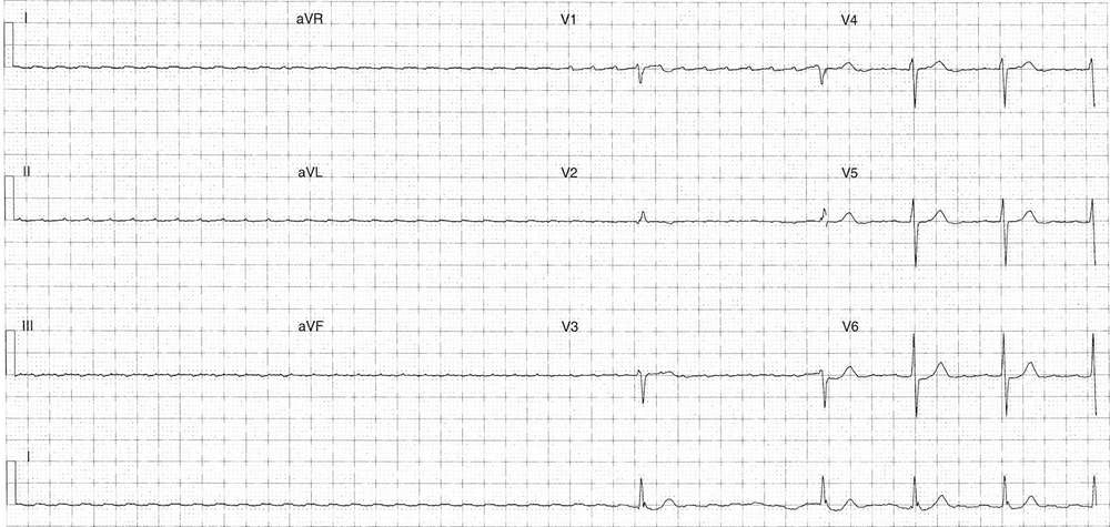
Key features
- Minimum 5.72 sec ventricular pause
- Evidence of atypical flutter waves
- Rate ~290 bpm
- Low voltage
- Positive in lead V1
- Following pause initiation of ventricular activity with increasing rate
- Ventricular 72 bpm (4:1 block) prior to end of ECG tracing
- QRS morphology sames as ECG above
What happened next ?
The patient’s heart rate rapidly returned to continued in a narrow complex tachycardia as per the first ECG. The patient underwent DCCV under procedural sedation and reverted following a single shock, post cardioversion ECG showed unremarkable normal sinus rhythm.
READ MORE: Atrial Flutter: ECG Interpretation [With Examples]
SIMILAR CASES: