This post is an answer to the ECG Case 312
- Rate: ~125 bpm
- Rhythm: regular sinus rhythm
- Axis: Normal
- Intervals:
- PR – Normal (~180ms)
- QRS – Normal (60ms)
- Segments:
- ST Elevation in leads: I (0.5-1mm), aVL (1mm), V1 (1mm), V2 (8-9mm), V3 (4mm), V4 (1mm)
- ST Depression in leads: II, III, aVF, V5-6
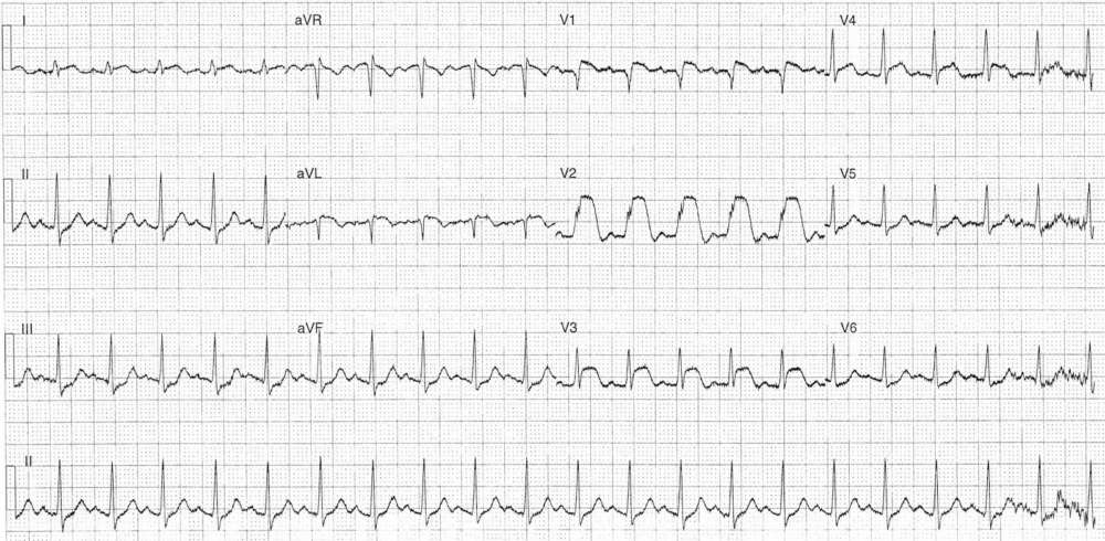
Interpretation
Anterior STEMI
What happened next ?
The patient was transferred for urgent coronography which showed:
- Left main 40% ostial lesion
- LAD diffuse disease with severe mid disease and severe distal disease – PCI with DES x 2
- Cx severe ostial
- RCA Diffuse mild-mod disease
Post procedure echo:
- Mod-severe segmental dysfunction with extensive anterior wall akinesis – EF 37%
- No significant valvular dysfunction
The patient had an uneventful further in-patient stay.
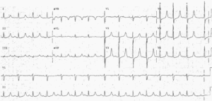
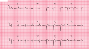
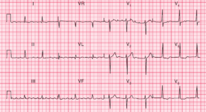
SIMILAR CASES: