This post is an answer to the ECG Case 314
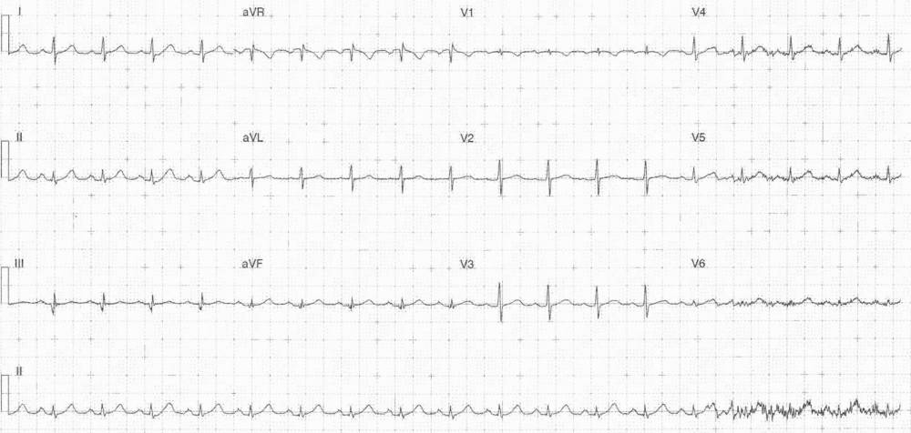
- Rate: 108 bpm
- Rhythm: Regular sinus rhythm
- Axis:Normal
- Intervals:
- PR – Normal (160-180ms)
- QRS – Normal
- QT – 300ms
- Segments:
- Up-sloping ST segment in lateral precordial leads
- Additional:
- Prominent T waves in leads II, aVF, V6 (in relation to QRS magnitude)
- Low QRS voltage
- No electrical alternans
- Baseline artifact affecting leads V4-6 and end of rhythm strip
Interpretation
- Prominent T waves in the setting of chest pain are concerning for OMI
- Requires serial ECG’s looking for progressive ST segment change
- Combination of low voltage and tachycardia should prompt consideration of pericardial effusion as a cause for the chest pain.
- As with all ECG’s the ECG features need to be considered in the patient’s specific clinical context
What happened next ?
The patient was admitted for investigation under the cardiology team. Serial troponins and D-dimer were negative and an angiogram showed only minor vessel irregularities.
READ ALSO: ECG Interpretation – All you need to know