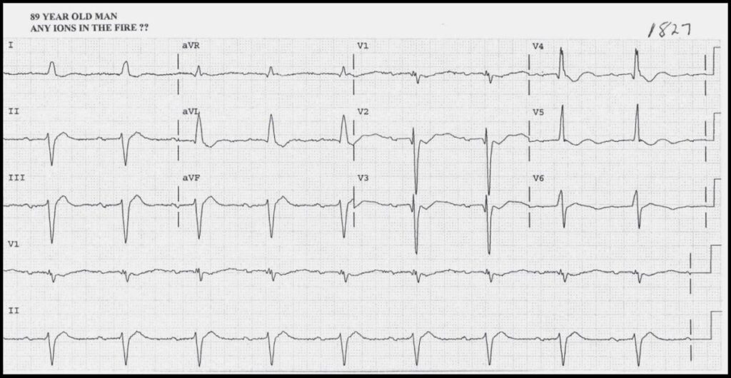
- The slow sinus P waves are conducted with a Prolonged PR Interval (can be caused by hypokalemia)
- There is Left Axis Deviation and probably LBBB (although V6 doesn’t look like LBBB)
- Q waves in V1 – V4 and slow R wave progression (could indicate old anterior MI)
- Prominent U waves in V2 – V5 (seen in hypokalemia)
- Shot QT Interval best seen in lateral and inferior leads (seen in hypercalcemia)
This man had widely metastatic prostate cancer, leading to Hypercalcemia.
Repetitive vomiting has resulted in Hypokalemia.
Read More about:
