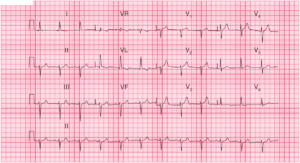
ECG Interpretation
- Sinus rhythm, rate 48/min
- Normal axis
- Small R waves in leads V2–V4 and a normal (tall) R wave in lead V5

Clinical interpretation
The small R waves in leads V2–V4 and the ‘sudden’ appearance of a normal R wave in lead V5 is called ‘poor R wave progression’, and despite the absence of Q waves this probably indicates an old anterior infarction. An alternative explanation might be poor lead positioning.
What to do ?
The ECG should be repeated, to ensure proper positioning of the chest leads. A serial troponins should be obtained to see if there’s acute MI. An echocardiogram, and a chest X-ray are needed, to see if left ventricular impairment is responsible for the breathlessness, and stress echocardiography or perfusion imaging are needed, to investigate the chest pain.
READ MORE about ECG Interpretation: All you need to know