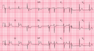
ECG Interpretation
- Ectopic atrial rhythm, with inverted P waves in leads II, III, VF, V3–V6; ventricular rate 69/min
- Normal axis
- Normal QRS complexes and T waves

Clinical Interpretation
This appears to be a stable rhythm originating in the atrial muscle rather than the SA node – hence the abnormal inverted P wave and the slightly short PR interval (130 ms).
This rhythm is not uncommon, and is usually of no clinical significance. It is unlikely to be the cause of her symptoms unless at times she has a paroxysmal atrial tachycardia.
What to do next?
Take a careful history and attempt to determine whether her symptoms sound like a paroxysmal tachycardia – ask about any sudden onset and ending of the palpitations; associated symptoms like breathlessness; precipitating and terminating factors; and so on. If in doubt, some sort of ambulatory recording will be needed.
READ MORE about: ECG Interpretation – All you need to know