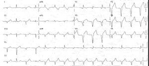
There is a regular rhythm at a rate of 140 bpm. The QRS complex duration is increased (0.12 sec). There is an RSR′ morphology in lead V1 (←) and an S wave in leads I and V4-V6 (←), diagnostic for a right bundle branch block (RBBB).
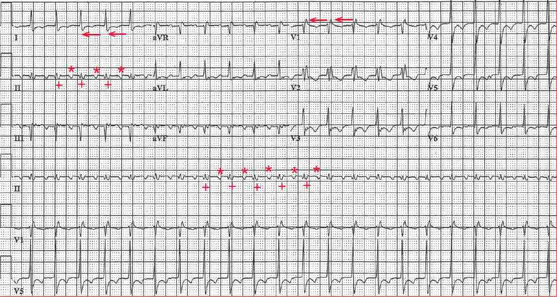
Negative atrial waveforms can be seen in leads II and aVF (*). A second atrial waveform (+) is seen at the end of the QRS complex, particularly in lead II. Although it looks like an S wave, it has the same morphology as the atrial waveform that is seen before the QRS complex and the interval between the two atrial waveforms is the same and stable, at a rate of 280 bpm.
The regular atrial rate of 280 bpm is diagnostic for atrial flutter, and there is 2:1 AV conduction. Atrial flutter is often a difficult rhythm to diagnose because one of the two flutter waves may be within the QRS complex, at the end of the QRS complex (resembling an S wave or even ST-segment depression), or at the beginning of the QRS complex (suggesting a Q wave).