This post is an answer to the ECG Case 223
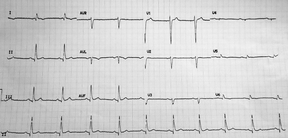
- Rate: 60 bpm
- Rhythm:
- Sinus rhythm
- Sinus arrhythmia
- Axis: Normal
- Intervals:
- PR – Normal (~200ms)
- QRS – Prolonged (120ms)
- QT – 440ms (QTc Bazette ~ 430 ms)
- Segments:
- ST Elevation V1 (<1mm)
- Additional:
- Lead I shows P and T wave inversion
- Lead aVR inverted P, QRS, and T wave
- Lead V1 higher overall voltage compared with other leads
- Lead V1 initial prominent R wave
- Leads V3-6 low voltage
- T wave inversion leads V5-6
Interpretation
Negative P wave in leads I and aVL with completely negative lead aVL should prompt consideration of LA/RA lead reversal. The low voltage lateral precordial leads, in conjunction with suspected lead reversal, should prompt consideration of dextrocardia. I only have limited information on this case but the patient had known dextrocardia.
Dextrocardia and the ECG
ECG features of dextrocardia are similar to a LA/RA lead reversal with the addition of precordial lead changes.
A LA/RA lead reversal results in the following ECG changes:
- Lead I becomes inverted
- Leads II and III switch places
- Leads aVR and aVL switch places
Unlike a lead reversal dextrocardia should manifest changes in the precordial leads which may include:
- Absent R wave progression
- Low voltage lateral precordial leads
- Leads V1 and V2 switch places
Interestingly in this ECG our patient has a normal axis, if the ECG was repeated with lead positions moved to account for the dextrocardia i.e RA lead on the left, LA lead on the right, and full right sided precordial leads (V1R-V6R) the resultant ECG would show a right axis deviation.
An important clue to spotting a lead reversal (true or apparent) in a patient with an abnormal axis is the P and T wave morphology; in this ECG both leads I and aVR have P and T wave inversion.
SIMILAR CASES ECG Case 68: Dextrocardia