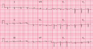
ECG Interpretation
- Sinus rhythm, rate 70/min
- Inverted P and T waves in leads I and aVL
- Right axis deviation
- Dominant R waves in lead VR
- No R wave development in the chest leads, with lead V6 still showing a right ventricular pattern
- Normal-width QRS complexes

Clinical Interpretation
This is dextrocardia. A normal trace would be obtained with the limb leads reversed and the chest leads attached in the usual rib spaces but on the right side of the chest.
What to do
Ensure that the leads are properly attached – for example, inverted P waves in lead I will be seen if the right and left arm attachments are reversed. Of course, this would not affect the appearance of the ECG in the chest leads.
READ MORE about ECG Interpretation: All you need to know