This post is an answer to the ECG Case 235
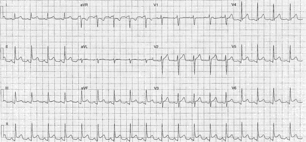
- Rate: 110 bpm
- Rhythm: Regular Sinus rhythm
- Axis: Normal
- Intervals:
- PR – Normal (160ms)
- QRS – Normal (80ms)
- QT – 300ms (QTc Bazette 410 ms)
- Segments:
- Widespread ST elevation in leads I, II, III, aVF, V2-6
- Concave morphology
- ST Depression in lead aVR
- Widespread ST elevation in leads I, II, III, aVF, V2-6
- Additional:
- PR depression in leads I, II, III, aVF, V4-6
- PR elevation in lead aVR
- Down-sloping T-P segment best seen in lead II
Interpretation
Pericarditis. Note sinus tachycardia, possible effusion ?
What happened next ?
The patient was admitted under the cardiology team. Blood tests showed a negative troponin but raised inflammatory markers and D-dimer.
A subsequent CTPA showed a pericardial effusion and the patient underwent pericardiocentesis for a large effusion, total drainage of ~900mls of fluid. The ultimate diagnosis was of viral pericarditis complicated by pericardial effusion.
READ MORE: Know the Differential for ST Segment Elevation: It’s More Than Just ACS
SIMILAR CASES:


