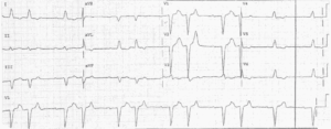
This post is an answer to the ECG Case 275
- Rate: 66 bpm
- Rhythm:
- Regular
- No P waves present
- Accelerated Idioventricular Rhythm (AIVR)
- Axis: Normal
- Intervals: QRS – Prolonged (160ms)
- Segments:
- Discordant ST segment changes
- Excessive depression in lead V5 and excessive elevation V3 (just on -0.25 ST elevation / QRS depth)
- Additional:
- LBBB Morphology
- Deep S in V1-3
- Broad R wave in lateral leads
- T waves massively disproportionate and peaked
- Note in leads V5-6 terminal portion of T wave becomes positive
- LBBB Morphology
The key abnormalities on this ECG are:
- AIVR
- LBBB with abnormal ST changes
- Massive peaked T waves
Broad differentials would include:
- Ischaemia
- Drug toxicity
- Acid-base disturbance
- Electrolyte abnormality
The diagnosis was hyperkalemia, urgent VBG was taken – K 8.8 mmol/L !
SIMILAR CASES: