This article is an answer to the ECG Case 174
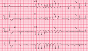
The ECG shows a regular rhythm at a rate of 90 bpm. There are no P waves seen in any of the leads. The QRS complex is very wide (0.24 sec), best established by measuring the QRS complex duration in lead V1 where the beginning and end of the QRS complex (J point) can be seen (└┘). The only condition associated with a QRS complex that is ≥ 0.24 second is hyperkalemia.
In general a bundle branch block or a ventricular complex will not cause a QRS complex to be this wide. A very wide QRS complex (up to 0.22 sec) may be seen with a severe dilated cardiomyopathy and this is a result of diffuse fibrosis and slowing of impulse conduction. In addition, the T waves are symmetric (upstroke and downstroke equal) (┴), which further supports hyperkalemia as the etiology.
The QRS complex axis is indeterminate between –90° and +/–180°. A wide QRS complex associated with an indeterminate axis occurs whenever there is direct stimulation of the ventricular myocardium and not when there is ventricular activation via the normal His-Purkinje system. This includes ventricular complexes, paced complexes or QRS complexes with a Wolff-Parkinson-White (WPW) pattern.
In addition, there are irregularities of the ST-T waves (^), best seen in lead V1. This finding, along with the indeterminate axis, suggests a diagnosis of ventricular tachycardia, along with hyperkalemia. This diagnosis is difficult to establish as the shift in the axis could be the result of the hyperkalemia.
Laboratory data indicated that he had a hyperosmotic hyperglycemic state accounting for the coma (glucose 800 mg/dL, serum osmolarity 4 00 mOsm/kg, pH 7.35 and no anion gap, and serum K 8.2 mmol/L).
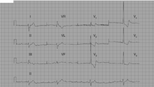
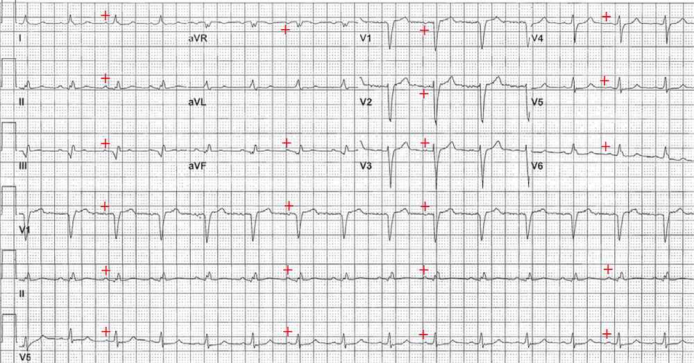
The next ECG was obtained after therapy for hyperkalemia was given and the potassium level corrected. There is a regular rhythm at a rate of 90 bpm. There is a P wave (+) before each QRS complex with a stable PR interval (0.22 sec). The P wave is positive in leads I, II, aVF, and V4–V6. Hence this is a normal sinus rhythm with a first-degree AV block. The QRS complex duration is normal (0.08 sec) as is the morphology.
READ MORE: ECG Interpretation: All you need to know
SIMILAR CASE: ECG Case 151: Hyperkalemia with Sine Wave Pattern