This post is an answer to the ECG Case 282
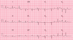
- Rate: 60 bpm
- Rhythm:
- Regular
- A-paced rhythm
- Atrial pacing spike with subsequent atrial depolarization P wave
- QRS Complexes conducted in native pattern via AV node
- Axis: Normal
- Intervals:
- PR – Normal (~200ms)
- QRS – Normal (100ms)
- QT – 440ms (QTc Bazette 440 ms)
- Segments:
- ST Elevation <1mm in lead aVR
- ST Depression in leads II, V4-6
- Additional:
- Biphasic T wave in lead V3
- T wave inversion in leads I, aVL, V4-6
- Borderline LVH by voltage criteria
Interpretation
ST Segment changes in lateral/high lateral leads.
Differentials
- ACS
- T wave memory
- Potential for ST / T wave changes to be due to a period of V-paced rhythm
- Secondary to LVH
- Drug effects especially digoxin although not typical appearance
What happened next ?
The patient was admitted under the Rehab team and had a troponin raise, following discussion with cardiology the patient was deemed for medical management only.
READ MORE: ECG Interpretation: All you need to know