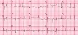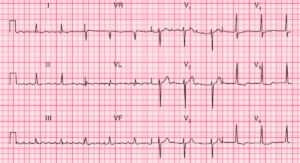
This post is an answer to the ECG Case 352
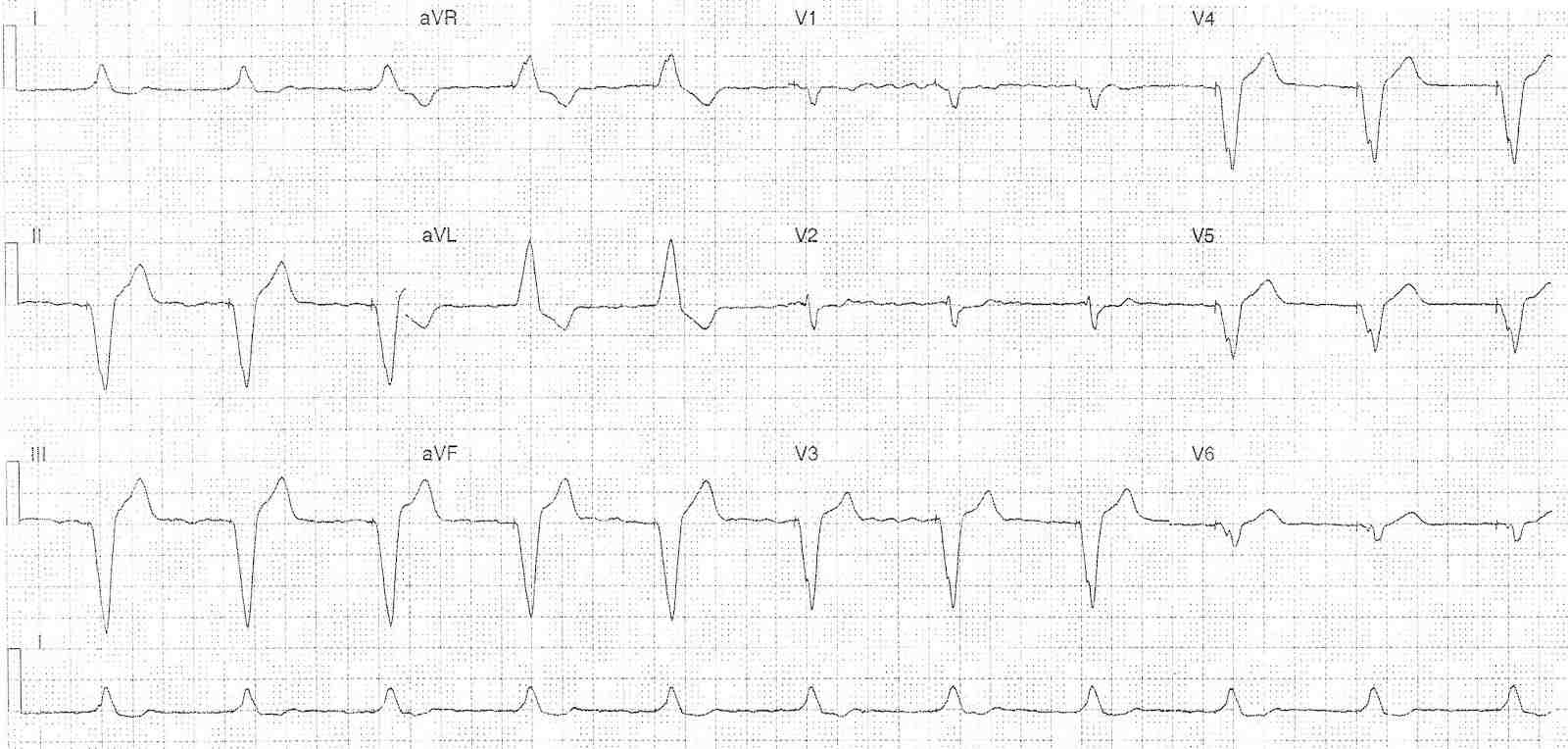
- Rate: 66 bpm
- Rhythm:
- No p waves
- Fibrillation waves best seen leads V1 and V3
- Regular ventricular rhythm
- Each ventricular complex associated with a pacing spike
- Best seen in leads II, aVR, aVF, V1, V3-6
- Axis: Lefts, superiorly at ~ -70 degrees
- Intervals: QRS – Prolonged (200ms)
- Additional:
- Appropriate discordant T wave and ST changes
- No evidence of excessive discordance
- Note QRS relatively low voltage in leads V1-2
- Need to check ECG lead placement
- Compare with old ECGs
- Monitor for dynamic change
Interpretation
- Ventricular paced rhythm
- All complexes paced
- Absence of native rhythm
- Lead placement: likely right ventricular apex
- No atrial activity seen
READ ALSO: ECG Interpretation: All you need to know