This post is an answer to the Case – 21-year-old Man with Exertional Dyspnea
Findings
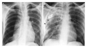
- On the frontal radiograph, a soft-tissue-density mass (arrow in Fig. 1) silhouettes the aortic arch and aorticopulmonary window. The left hilar vessels (asterisk in Fig. 1) can be seen through the mass (hilum overlay sign) and the descending thoracic aortic line is preserved.
- On the lateral, the mass fills in the retrosternal clear space. The trachea is also displaced posteriorly and compressed against the spine.
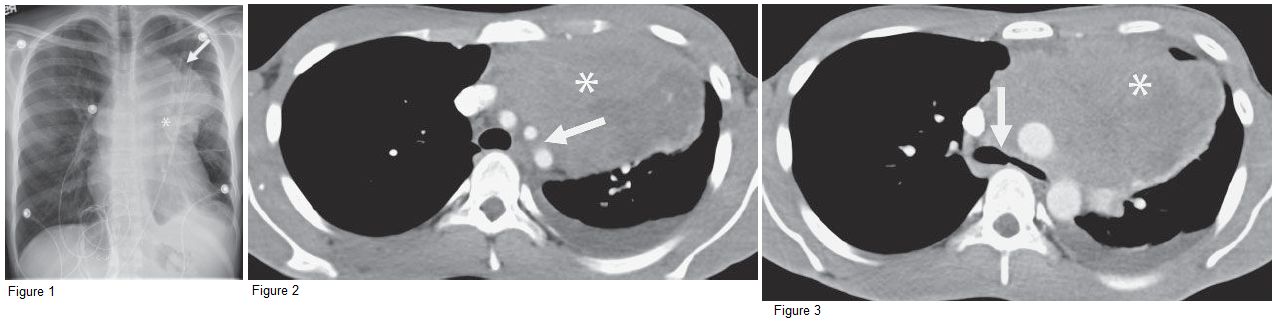
- CT images demonstrate a slightly heterogenous soft tissue mass (asterisk in Figs. 2 and 3) centered within the anterior mediastinum, encasing the great vessels (arrow in Fig. 2) and displacing the trachea and left mainstem bronchus posteriorly (arrow in Fig. 3).
Differential Diagnosis
Because the mass is mostly soft tissue with only few areas of fluid attenuation, the differential diagnosis would include lymphoma, malignant germ cell tumor, and less likely thymic mass, such as thymoma.
Teaching Points
- The most commonly encountered anterior mediastinal masses in adults are thymoma and lymphoma. Germ cell tumors are more common in younger patients.
- While Hodgkin’s lymphoma is the most common form presenting in the anterior mediastinum, diffuse large B-cell lymphomas have a propensity for the anterior mediastinum in young adults and can present with symptoms related to compression of vessels and the airway.
- Lymphomas of the mediastinum tend to surround vessels but rarely compress them. Occasionally, lymphomas extend anteriorly into the chest wall.
- The hilum overlay sign (lack of obscuration of the hilar structures [i.e. bronchi, pulmonary arteries and veins]) on plain radiograph indicates that a mass lies within the anterior or posterior mediastinum. When the posterior mediastinal lines, descending thoracic aortic stripe, and paravertebral lines are preserved, localization can be made to the anterior mediastinum.
Management
If no easily accessible nodes, such as supraclavicular or axillary, are involved, many anterior mediastinal masses can be approached parasternally via CT or US guidance for tissue sampling. For lymphoma, fine-needle aspiration for flow cytometry and core biopsy should be performed.
Further Reading
Whitten CR, Khan S, Munneke GJ, Grubnic S. A diagnostic approach to mediastinal abnormalities. Radiographics. 2007 May-Jun;27(3):657-71. doi: 10.1148/rg.273065136. PMID: 17495284.