This post is an answer to the Case – Severe Epigastric Pain
What abnormality can you see ?
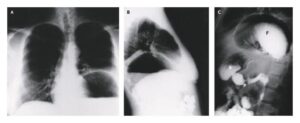
- Left Image: ‘Free gas under diaphragm’ – indicates free peritoneal gas between the dome of the hemidiaphragm and superior surface of liver.
- Right Image: Supine abdominal radiograph showing signs of extraluminal gas including the Rigler’s sign (also known as double wall sign) and triangular sign.
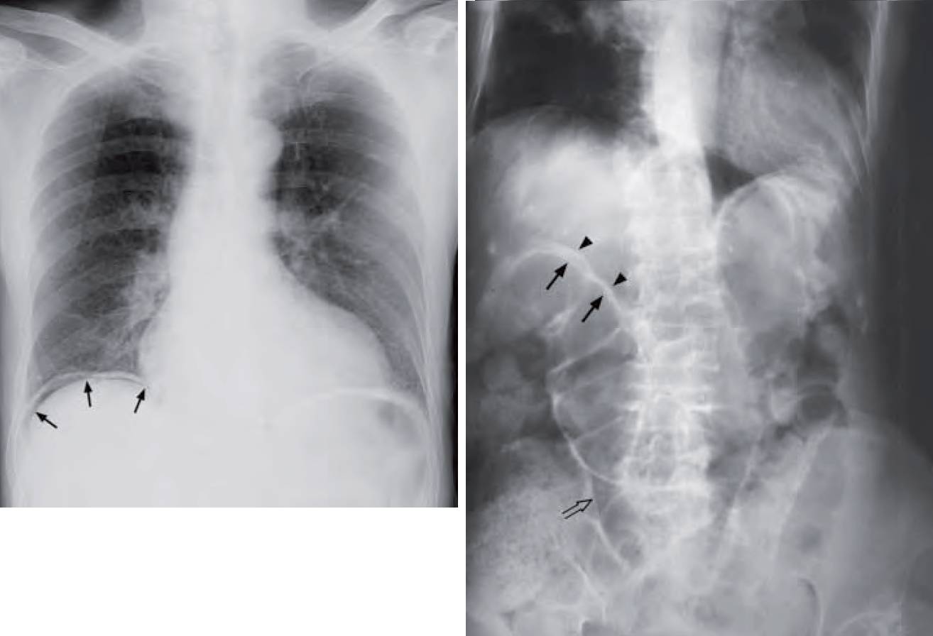
Right Image: Rigler’s sign (also known as the double wall sign) – the inner mucosal surface of the bowel wall is delineated by intraluminal air (arrows) and the outer serosal surface of the bowel wall is also delineated by extraluminal intraperitoneal free air (arrow heads). Note the triangular collection of gas between adjacent loops of bowel (open arrow) – triangular sign.
Diagnosis: Pneumoperitoneum secondary to perforated peptic ulcer
Discussion
Pneumoperitoneum (gas within the peritoneal cavity) may be due to many causes, including:
- Gastro-intestinal tract perforation (peptic ulcer, diverticulum, ischaemic bowel, etc.)
- Penetrating injury of the abdomen
- Pneumomediastinum with peritoneal extension
- Gas-forming intraperitoneal infection
- Air entering via the female genital tract
30% of patients with perforated peptic ulcer do not have CXR finding of pneumoperitoneum. An erect CXR is the initial imaging investigation of choice.
The patient should be kept in the erect position for at least 10 minutes before the radiograph is taken to ensure adequate time for free gas to collect in a nondependent portion of peritoneal cavity. If this is not possible, an AXR in the left lateral decubitus position (lying on the side with left side of body dependent) is an alternative. This way, any free gas will float to the nondependent part of the peritoneal cavity (i.e. right side). Pneumoperitoneum, even if substantial, on supine AXR is sometimes difficult to detect.
Radiographic signs include:
- Rigler’s sign – bowel wall outlined by intraluminal (normal) and peritoneal gas (abnormal)
- Triangular sign – gas collecting between adjacent bowel loops
- Football sign – gas in peritoneum within a distended abdomen outlining intraabdominal viscera (see figures in necrotising enterocolitis in paediatric section)
- Gas outlining the falciform ligament (see figures in necrotising enterocolitis in paediatric section)
If clinical suspicion remains high despite a negative CXR, a water soluble contrast examination of the bowel may help to confirm/rule out the diagnosis (by demonstrating contrast leakage from the site of perforation
A CT scan is better at demonstrating the extent of intraperitoneal free gas and detecting small amounts of gas. Note that hepatic interposition of the bowel may mimic free gas under the diaphragm. This is called Chilaiditi’s sign and is seen in asymptomatic patients. When it is associated with symptoms, either intermittent or persistent, it is known as Chilaiditi’s syndrome.
SIMILAR CASES: