
This post is an answer to the Case – Severe Sore Throat, Painful Swallowing and Fever
Interpretation
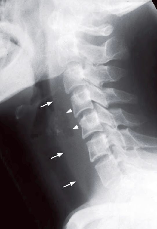
- Widened retropharyngeal soft tissues
- A localized gas locule in the retropharyngeal space
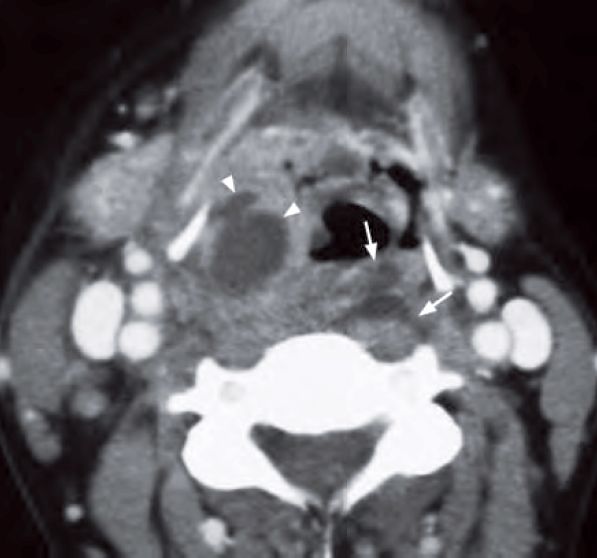
A contrast enhanced CT neck was performed (image below) which showed:
- Prominent retropharyngeal soft tissues
- Large rim-enhancing lesions with necrotic centre in retropharyngeal soft tissue consistent with an abscess
Diagnosis
Lateral radiograph of the neck shows thickening of the retropharyngeal soft tissues (arrows) and localized gas lucency within (arrowheads) suggestive of a retropharyngeal abscess.
Key Points
- Gas locules within widened retropharyngeal soft tissues on plain radiographs in the appropriate clinical setting is highly suggestive of retropharyngeal abscess.
- CT is the imaging modality of choice for exact anatomical localization and extent of the abscess.