This post is an answer to the Case – Patient with Colicky Abdominal Pain
What radiological abnormalities can you identify on this radiograph ?
- There are multiple dilated bowel loops roughly central abdominal in location.
- Valvulae conniventes appearing as striations crossing completely across the width of the bowel loops.
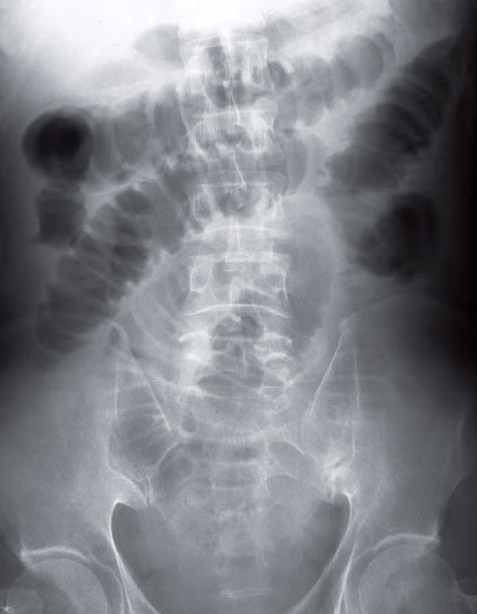
Diagnosis: Small Bowel Obstruction
Role of imaging in the evaluation of bowel obstruction
- To confirm the diagnosis of bowel obstruction
- To localize the site/level of obstruction
- To detect obstructing lesions like adhesions, tumours, volvulus, hernia, etc.
- To detect possible complications such as strangulation and bowel ischaemia/ infarct
READ MORE: Abdominal Pain in the Emergency Department
SIMILAR CASES: