This article is an answer to the Case – Ulcer and Subcutaneous Nodules
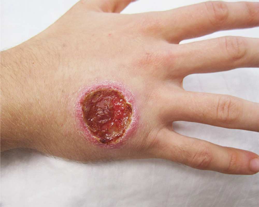
A punch-biopsy specimen of the edge of the ulcer showed a dense infiltrate of lymphocytes and histiocytes containing intracellular organisms consistent with leishmania amastigotes.
A polymerase-chain-reaction assay of the specimen was performed, and the species was identified as Leishmania panamensis. Treatment with oral miltefosine (at a dose of 50 mg three times daily for 28 days) was initiated.
Cutaneous leishmaniasis caused by L. panamensis can disseminate to the oronasopharyngeal mucosa. A fiberoptic examination of the nasopharynx performed 6 days after the patient began treatment showed no evidence of mucosal disease.
Ten days after the completion of therapy, the ulcer had healed with an atrophic scar, and the number and size of the subcutaneous nodules were markedly reduced.
References
- Sara Rahimi, Javad Rafinejad, Amir Ahmad Akhavan, Reza Ahmadkhaniha, Mahmood Bakhtiyari, Ali Khamesipour, Kamran Akbarzadeh. (2023) The therapeutic effect of larval saliva and hemolymph of Lucilia sericata on the treatment of Leishmania major lesion in BALB/c mice946. Parasites & Vectors 16:1.
- Mehdi Ghazanfari, Bahador Shahriari, Vahid Rahnama, Meisam Khazaei, Shahrbanou Naderi, Mohammad Hossein Motazedian. (2022) The level of interleukin-17, 23, and gamma interferon in cutaneous leishmaniasis patients before and after intra lesion treatment. Journal of Parasitic Diseases 46:2, 476-482.
- Boniface P. Kamdem, Ferreira I. Elizabeth. (2021) The Role of Nitro (NO2-), Chloro (Cl), and Fluoro (F) Substitution in the Design of Antileishmanial and Antichagasic Compounds. Current Drug Targets 22:4, 379-398.