
A 51-year-old man presented with fever and general malaise of 2 weeks’ duration. He had had diabetes mellitus for the preceding 20 years, and at the time of presentation this condition was poorly controlled.
On admission to the hospital, his white-cell count was 10,800 per cubic millimeter (normal range, 3900 to 9800). The C-reactive protein level was 8.6 mg per deciliter, blood urea nitrogen 90 mg per deciliter (32 mmol per liter), creatinine 4.9 mg per deciliter (430 μmol per liter), and glycated hemoglobin 11.2%.
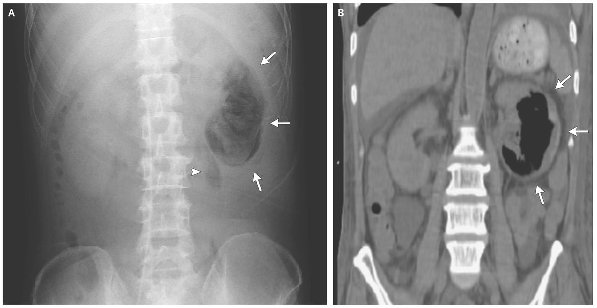
Abdominal radiography (Panel A) and computed tomography (Panel B) revealed gas collection in the parenchyma (arrows) and perinephric space (arrowhead) of the left kidney.
The patient received a diagnosis of emphysematous pyelonephritis, possibly caused by retrograde infection related to diabetes-associated neurogenic bladder.
The greatest risk factors for development of emphysematous pyelonephritis are poorly controlled diabetes mellitus and ureteric obstruction.
Escherichia coli was isolated in blood culture but not in urine cultures. The patient was treated with antibiotics; the infection resolved, and renal function improved (the blood urea nitrogen level declined to 23 mg per deciliter [8 mmol per liter] and creatinine to 2.6 ml per deciliter [230 μmol per liter]).
SIMILAR CASE: Acute Emphysematous Cystitis with Air in the Urinary Tract