This article is an answer to the Case – Left-Sided Chest Mass
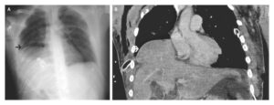
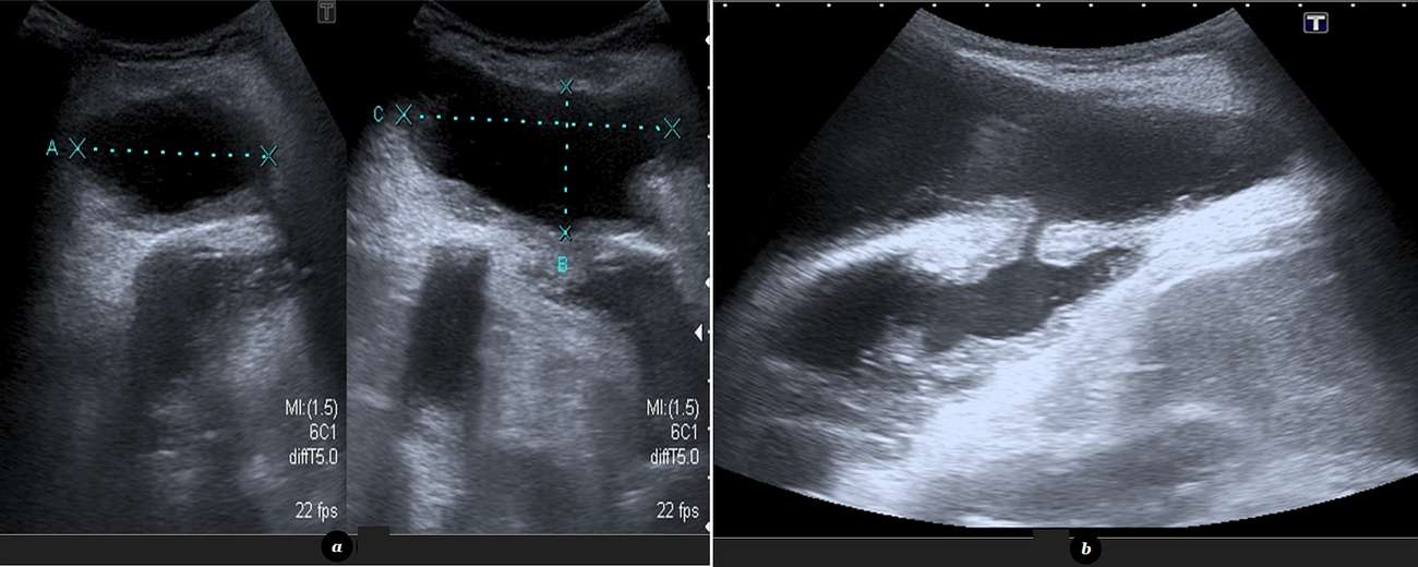
Chest radiography demonstrated a loculated left pleural effusion with associated soft-tissue opacity in the left chest wall. An abdominal ultrasound (US) was performed in order to better characterize the radiographic findings, showing a subcutaneous fluid collection with fine internal debris, measuring 5 x 3 x 7 cm, that communicated with the pleural cavity through a well-defined tract (image below).
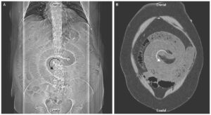
Imaging findings were further investigated with contrast-enhanced computed tomography (CT) imaging. CT revealed a loculated left pleural effusion with thickened, contrast-enhanced and partially calcified pleural surfaces that communicated with an organized, subcutaneous chest-wall fluid collection.
Both the subcutaneous collection and the pleural effusion were drained and the fluid samples from both cavities were analyzed: they were positive for acid fast bacilli and cultures confirmed the presence of Mycobacterium tuberculosis. Anti-bacillary treatment was initiated and the patient was discharged totally asymptomatic. Consultation with the thoracic surgeon was scheduled.
Discussion
Empyema necessitans (or empyema necessitatis) is a complication of pulmonary infection, most frequently secondary to Mycobacterium tuberculosis infection reactivation, although it can also occur with actinomycosis and pyogenic bacterial infection [1, 2]. It represents extension of pus from the pleural cavity to the chest wall (most frequently), but also to the bronchi, oesophagus, breasts or retroperitoneum [3, 4].
The patients generally present with an enlarging, occasionally painful, erythematous chest wall mass, most frequently affecting the second through sixth intercostals spaces [5]. Associated respiratory symptoms such as cough or dyspnoea and pleuritic-type chest pain also occur.
While chest radiography can suggest the diagnosis, demonstrating a loculated pleural effusion in a patient with past history of pulmonary tuberculosis presenting with an anterior chest wall mass, the definitive imaging findings are usually seen with CT. Contrast-enhanced CT clearly demonstrates the existence of a communication between the empyema (i.e.: a loculated pleural effusion with thickened and enhancing pleural surfaces) and a well-deliniated chest wall fluid collection with thickened and enhancing walls, representing an abscess [6]. US and CT can also aid in the therapeutic process, guiding the thoracostomy necessary to drain the pleural cavity.
Generally there is a good response to tube-thoracostomy and parenteral antibiotic therapy but, in some cases, there is necessity to perform a thoracotomy and pleural decortication to definitely solve the problem [4]. Tuberculous empyema necessitans has a good clinical outcome when managed accurately and promptly, with an associated mortality rate less than 5% [7]. Accurate diagnosis based on imaging evaluation and adequate antibiotic therapy are crucial for management of this disease.
References
- Freeman AF, Ben-Ami T, Shulman ST (2004) Streptococcus pneumoniae empyema necessitatis. Pediatr Infect Dis J 23: 177-9 (PMID: 14872190)
- Soto-Hurtado EJ, Marin-Gamez E, Segura-Dominguez N, Jimenez-Onate F (2005) Pleural aspergillosis with bronchopleurocutaneous fistula and costal bone destruction: a case report. Lung 183: 417-23 (PMID: 16465601)
- Glicklich M, Mendelson DS, Gendal ES, Teirstein AS (1990) Tuberculous empyema necessitatis: computed tomography findings. Clin Imaging 14:23-25 (PMID: 2322879)
- Mirza B, Ijaz L, Sheikh A (2011) A rare presentation of empyema necessitatis. Lung India 28(1): 73–74 (PMID: 21654996)
- Kellie SP, Shaib F, Forster D, Mehta JP (2010) Empyema Necessitatis. Chest 138:39A
- Choi J \”et al\” (2001) CT Manifestations of Late Sequelae in Patients with Tuberculous Pleuritis. American Journal of Roentgenology 176: 441-445 (PMID: 11159091)
- Porcel JM, Madronero AB, Bielsa S (2004) Tuberculous empyema necessitatis. Respiration 71(2):191 (PMID: 15031577)