This article is an answer to the Case – A 6-year-old Boy with Several Hours of Vomiting and Dyspnea
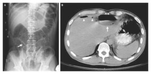
A postoperative anteroposterior plain radiograph (Panel A) showed a raised left hemidiaphragm (arrow) and a left chest tube (arrowhead) that had been placed earlier to drain a left hemothorax. The boy was discharged from the hospital after recovering from his injuries.

At the time of the current presentation, physical examination revealed no breath sounds over the left hemithorax. Anteroposterior plain radiography of the chest (Panel B) showed a large, gas-filled structure in the left hemithorax (long arrow), near-complete collapse of the left lung (short arrow), and a marked rightward shift of the mediastinum (arrowhead).
The left hemidiaphragm could not be clearly seen. After the administration of contrast material through a nasogastric tube, repeat chest radiography was performed and confirmed that the stomach lay within the left hemithorax (Panel C).
The patient had a left posterolateral diaphragmatic rupture and underwent emergency surgery, during which the stomach, spleen, left kidney, and part of the transverse colon were repositioned within the abdomen and the rupture repaired.
A postoperative chest radiograph showed reexpansion of the left lung (Panel D). The patient had a smooth recovery and was discharged home 10 days after the surgery.