This article is an answer to the Case – Patient with Intermittent Dysphagia and Regurgitation
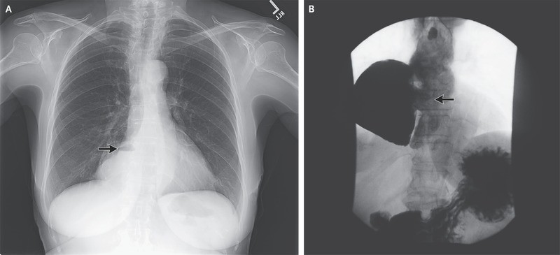
A chest radiograph showed a density containing an air–fluid level to the right of the heart (Panel A, arrow). Radiography of the esophagus revealed an epiphrenic diverticulum, 10 by 15 cm (Panel B, arrow), and delayed emptying at the gastroesophageal junction with esophageal dilatation and tortuosity, which are findings consistent with the patient’s diagnosis of achalasia.
Endoscopic examination confirmed the presence and position of the diverticular opening, and a yeast infection was confirmed by culture.
Because of the patient’s progressive symptoms, a laparoscopic resection of the diverticulum, Heller’s myotomy, and Dor’s fundoplication were performed, without complications.
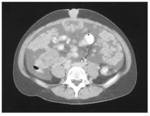
Epiphrenic diverticula typically arise just proximal to the lower esophageal sphincter. They are thought to be caused by ballooning of the esophageal mucosa and submucosa through a weakened area in the muscular layer as a result of increased intraluminal pressure, and they are commonly associated with an underlying motility disorder such as achalasia, as in this case.
Two years after surgery, the patient remained free of symptoms.