This article is an answer to the Case – Patient with 3-month History of Coughing Reddish Sputum, Weight Loss, and Fever
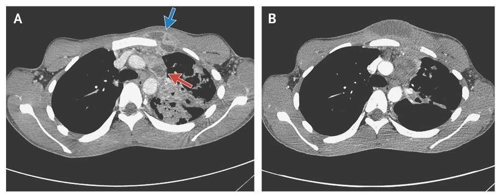
A computed tomographic (CT) scan of the chest obtained on admission showed a collar-button abscess (a subcutaneous abscess connected to a deeper abscess by a passage), consisting of an abscess in a lymph node in the left mediastinum (Panel A, red arrow) connected to a subcutaneous abscess in the anterior chest wall (Panel A, blue arrow). CT also revealed an extensive parenchymal abnormality in the left upper lobe.
A sputum smear showed acid-fast bacteria, and a culture grew pansusceptible Mycobacterium tuberculosis. Tests for human immunodeficiency virus were negative.
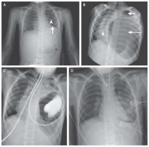
The patient received 8 weeks of standard antituberculous treatment, but the infection progressed, with increased swelling, erythema, and fluctuation of the subcutaneous abscess (Panels B). The abscess was subsequently treated with open drainage.
Pharmacologic treatment of a tuberculous abscess is seldom sufficient, and open drainage or surgical excision is often required. The patient has been without signs of relapse since surgery but required plastic surgery to improve the cosmetic result after drainage.