This article is an answer to the Case – Chest Pain, Anorexia, Weight Loss and Cachexia
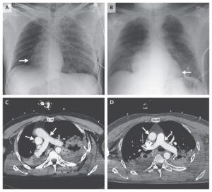
Upright posteroanterior and lateral chest radiographs showed a large well-marginated rounded mass in the posterior mediastinum.
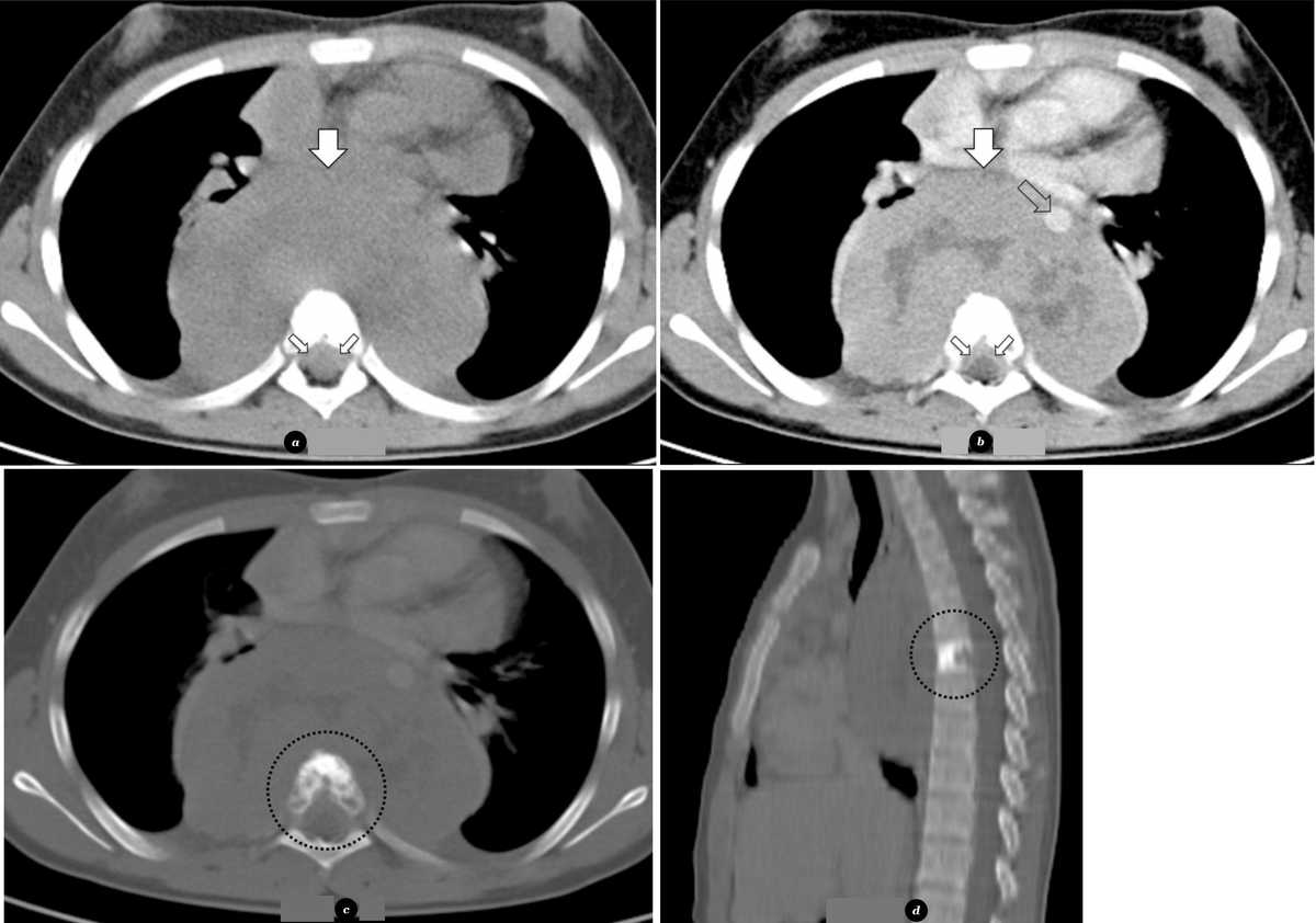
Chest CT revealed a midline well-defined soft-tissue mass with a diameter of 12 cm, located in the posterior mediastinum, adjacent to the vertebral bodies from T3 to T8, encasing the descending aorta (image below). The mass was associated with permeative destruction and osteosclerosis of the 6th thoracic vertebral body (image (d)). There was also extension to the spinal canal through the neural foramina at the 6th to 7th thoracic vertebrae.
Axial T1-weighted, T2-weighted and contrast-enhanced T1-weighted MR images confirmed extension to the spinal canal with involvement of epidural space, displacing the spinal cord posteriorly. Coronal and sagital T2-weighted MR images depicted cranial and caudal extent of the tumour, as well as the extension to the spinal canal though the neural foramina at the 6th to 7th thoracic vertebrae.
Cross-sectional imaging investigation for staging was negative for metastatic involvement.The patient underwent biopsy under thoracoscopy and the tumour was diagnosed as a Ewing’s sarcoma. The tumour was considered unresectable at initial presentation and chemotherapy was initiated in order to reduce its size and allow potential future complete tumour resection.