This article is an answer to the Case – Incidental Finding on Chest X-Ray
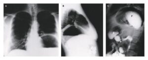
After acute coronary syndrome was ruled out, she was treated for gastroesophageal reflux, which relieved her symptoms. A chest radiograph showed a dense opacity in the upper area of the left lung.
The differential diagnosis for this abnormality includes:
- old calcified empyema
- hemothorax
- oleothorax
Given her history of treatment for tuberculosis, the most likely diagnosis was oleothorax — a treatment for pulmonary tuberculosis, abandoned long ago, that involved the instillation of oil into the pleural space to collapse the involved lung.
Typically, after treatment, which could last up to 2 years, the oil was aspirated. However, asymptomatic patients were sometimes lost to follow-up and the oil was left in place, as occurred in this patient.
Long-term complications, including superimposed infection and airway obstruction, have been reported. This patient had no complications or symptoms related to oleothorax.