This article is an answer to the Case – Patient with Acute Dyspnea and Chest Pain and a History of Peptic Ulcer
He had hypotension and tachycardia with an oxygen saturation of 92% while he was breathing 2 liters of oxygen per minute through a nasal cannula.
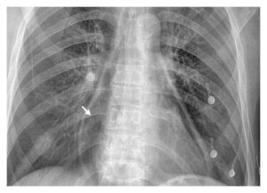
Chest radiography showed air in the pericardium (Panel A, arrow). Computed tomography of the chest confirmed the presence of a large pneumopericardium (Panel B, arrow) but no pneumothorax or pneumoperitoneum.
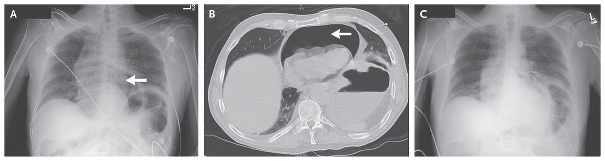
Emergency pericardiocentesis was performed, and a large amount of air was aspirated. The patient’s vital signs immediately stabilized. Repeat chest radiography showed improvement of the pneumopericardium (Panel C).
Further workup revealed that the patient had a penetrating peptic ulcer, with fistula formation from the fundal aspect of the stomach through the diaphragm and into the pericardial space. The fistula was ultimately repaired surgically.