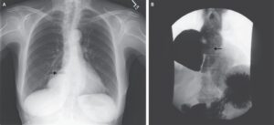
This post is an answer to the Case – Shortness of Breath After Falling Out of a Tree
Findings
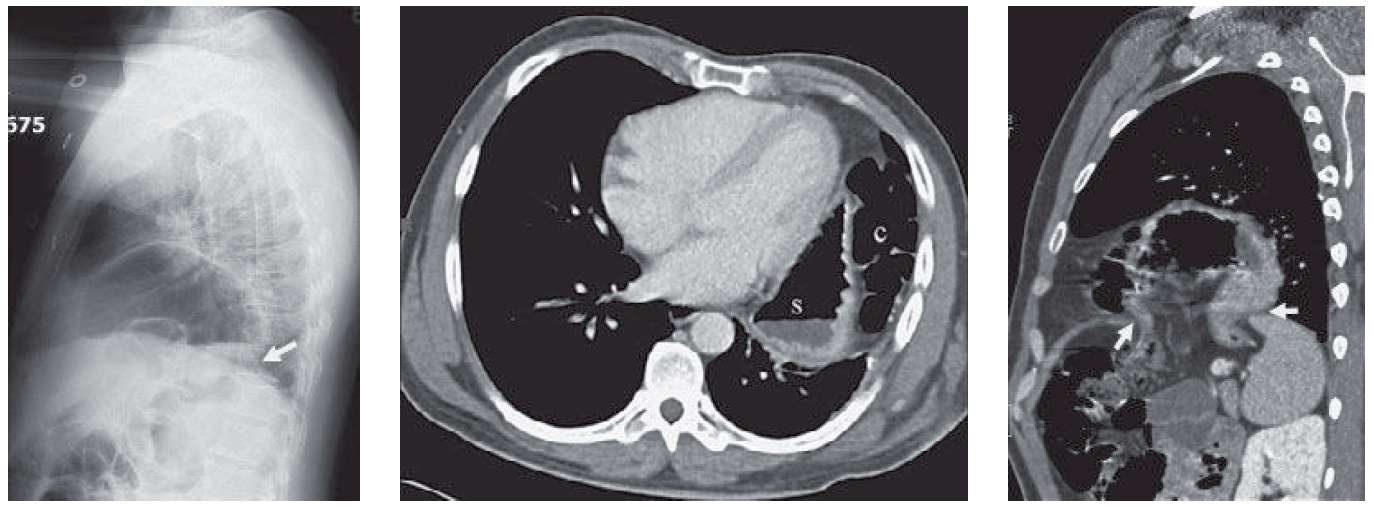
- Chest radiograph demonstrates elevation of the gastric bubble. The contour of the apparent diaphragm is irregular and the heart is shifted rightward.
- Lateral examination demonstrates a focal extrusion of the stomach above the level of the left hemidiaphragm with a neck (arrow on CXR).
- CT images demonstrate stomach (s) and colon (c) abutting the chest wall.
- Sagittal reconstruction demonstrates a large diaphragmatic defect (white arrows on the CT image) with herniation of the stomach, colon, and mesenteric vessels/fat into the thoracic cavity. There is a waist at the level of the herniation (collar sign).
Differential Diagnosis
Differential diagnosis for apparent elevation of the left hemidiaphragm consists of traumatic rupture, paralysis, or eventration. Shift of the heart rightward and the normal posterior sulcus would argue against paralysis. The acute angle between the stomach and left hemidiaphragm would favor traumatic rupture over eventration.
Teaching Points
- Traumatic rupture of the diaphragm is seen in <5 % of blunt trauma cases and is harder to image in penetrating trauma.
- Elevated abdominal pressures force abdominal organs into the pleural space. Positive-pressure ventilation may mask rupture by reducing the pleural pressure gradient and preventing herniation.
- Left -sided injuries are easier to identify on imaging.
- Elevated diaphragm in the setting of trauma should raise the suspicion of a diaphragmatic hernia.
- Chest radiograph findings include apparent diaphragmatic elevation, irregular or discontinuous diaphragmatic contour, abnormal course of a feeding tube, or gas-containing viscera above the diaphragm.
- MDCT findings include the collar sign, the dependent viscera sign (stomach resting on the posterior chest wall), herniated viscus or mesentery, large defect in the diaphragm, retraction of the injured part of the muscle adjacent to the tear, and the cottage bread sign (referring to a right injury with herniated liver simulating brioche).
Management
A diaphragmatic herniation requires prompt surgical attention due to the increased risk of strangulation of the herniated viscera.
Further Reading
Iochum S, Ludig T, Walter F, Sebbag H, Grosdidier G, Blum AG. Imaging of diaphragmatic injury: a diagnostic challenge? Radiographics. 2002 Oct;22 Spec No:S103-16; discussion S116-8. doi: 10.1148/radiographics.22.suppl_1.g02oc14s103. PMID: 12376604.