Laboratory evaluation showed leukocytosis, with a white-cell count of 20,400 per cubic millimeter and 82% segmented neutrophils. No anemia was found.
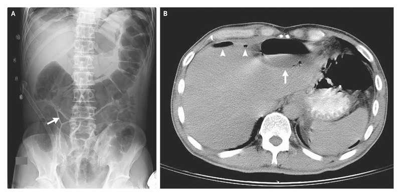
Supine plain radiography of the abdomen revealed a small triangular pocket of air outlined by three adjacent bowel loops (Panel A, arrow), a finding that was consistent with the presence of free intraperitoneal air; this is known as the telltale triangle sign.
Abdominal computed tomography confirmed the presence of pneumoperitoneum (Panel B, arrowheads) and also showed a subdiaphragmatic abscess with an air–fluid level (Panel B, arrow).
During surgery, a perforated gastric ulcer and two intraabdominal abscesses were found. The patient underwent ulcerectomy with pyloroplasty and drainage of the abscesses; he also received a proton-pump inhibitor. The patient was doing well at follow-up 6 months after surgery.


