This post is an answer to the Case – 73-year-old Patient with Pneumonia
Findings
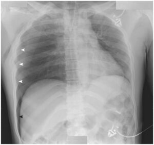
- Chest radiograph demonstrates a moderate-sized left pleural effusion.
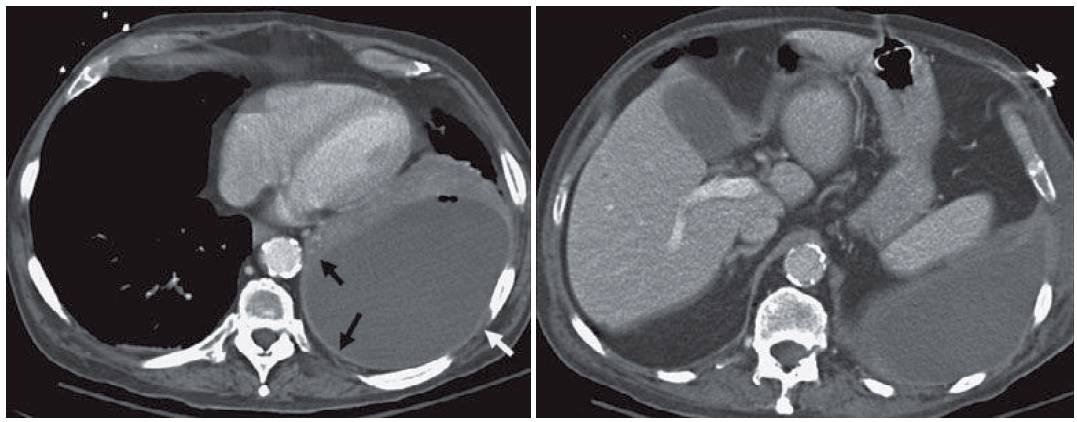
- CT demonstrates the effusion with bubbles of gas. Smooth thickening and enhancement of the visceral and parietal pleura consistent with a “split pleura” sign (black arrows) and subpleural edema (white arrow) are seen. The thickening is even easier to see on a more caudal image.
Differential Diagnosis
The differential diagnosis for a unilateral medium pleural effusion is lead by hemothorax, empyema, parapneumonic effusion, and malignancy. When pleural thickening is identified, malignancy and empyema become the major two considerations. The presence of subpleural edema favors empyema.
Teaching Points
- Empyema refers to pus in the pleural space. Most oft en this occurs in the setting of a pneumonia. Empyema connotes a fibropurulent phase, as opposed to a simple exudative parapneumonic effusion.
- The split pleura sign is indicative of a complex pleural effusion. In the setting of a smooth pleural surface one would favor an empyema in the differential.
- The split pleura sign is seen in about 50 % of cases of empyema. Isolated parietal pleura enhancement is also seen in about 50 % of cases.
- Nodular pleural thickening raises the concern for malignancy, such as mesothelioma or metastatic disease.
- An empyema can be distinguished from a lung abscess based on shape, as the empyema tends to be lenticular and assumes different dimensions in different axes. Pulmonary abscesses tend to be spherical, having nearly the same dimension in the sagittal, transverse, and coronal planes.
- Gas in the pleural space may be seen in the setting of an empyema, recent intervention, or bronchopleural fistula.
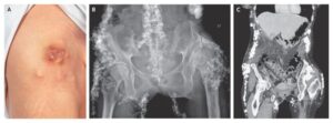
- Occasionally, an empyema can break through the chest wall. This is known as an empyema necessitans and can be seen in the setting of tuberculosis, nocardia, and actinomycosis empyema.
Management
- Initial management consists of obtaining cultures of the pleural fluid and placing a chest tube for evacuation.
- An empyema may become organized and create a rind that restricts lung expansion. This is known as a trapped lung and requires decortication.
SIMILAR CASES:
Further Reading
Kraus GJ. The split pleura sign. Radiology. 2007 Apr;243(1):297-8. doi: 10.1148/radiol.2431041658. PMID: 17392263.