This post is an answer to the Case – Inflammatory leg nodules
A man aged 42 years with alcohol use disorder was admitted to hospital for acute abdominal pain that started 12 h previously. Blood tests showed a high lipase concentration (745 IU/L), and an abdominal CT scan confirmed the diagnosis of acute pancreatitis with one acute necrotic collection, characterised as moderately severe acute pancreatitis according to the Atlanta classification.
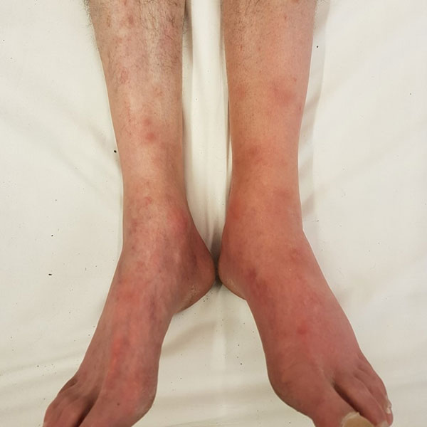
2 days later, the man presented with acute pain in both legs and local examination showed multiple purpuric erythematous macular lesions. On day 4, these lesions were characterised as subcutaneous erythematous nodules associated with inflammatory oedema of the left ankle although both legs were affected.
A punch biopsy was performed and histology showed cytosteatonecrosis in the superficial part of the hypoderm, characterised by lobular panniculitis with dense inflammation, and necrosis together with foaming macrophages and so-called ghost adipocytes (without nuclei). No lesions that would suggest vasculitis or T-cell lymphoma were reported.
Blood tests ruled out autoimmune disease or vasculitis and the concentration of α1-antitrypsin was normal. Bacteriological and mycobacteriological cultures were negative. Skin lesions, oedema, and pain in the lower limbs resolved spontaneously after 7 days, confirming the diagnosis of pancreatic panniculitis.
Systemic panniculitis is caused by the release of pancreatic enzymes, leading to necrosis of hypodermic fat tissue and sometimes bone tissue. Systemic panniculitis mainly affects men aged 40–70 years who have a history of chronic alcohol use disorder. Lesions are typically dermo-hypodermic inflammatory nodules or plaques, predominantly on the lower limbs and fingers. In case of chronic pancreatitis or pancreatic carcinoma, dermatological lesions might precede the usual manifestations of underlying pancreatic disease by several weeks to several months. Systemic symptoms can occur, such as fever, arthralgia, or bone pain due to intraosseous fat necrosis. Histologically, a predominantly lobular pattern with fat necrosis and ghost adipocytes is pathognomonic.
Treatment with glucocorticoids and non-steroidal anti-inflammatory drugs is usually ineffective. Apart from the usual spontaneous resolution of skin lesions with the treatment of pancreatitis, octreotide has been proposed as an option due to an inhibitory effect on lipase activity, and tetracycline has also been proposed in the treatment of panniculitis.
SIMILAR CASE: Epigastric Pain, Vomiting and Elevated Lipase Level