This article is an answer to the case – Infant with Poor Feeding and Vomiting
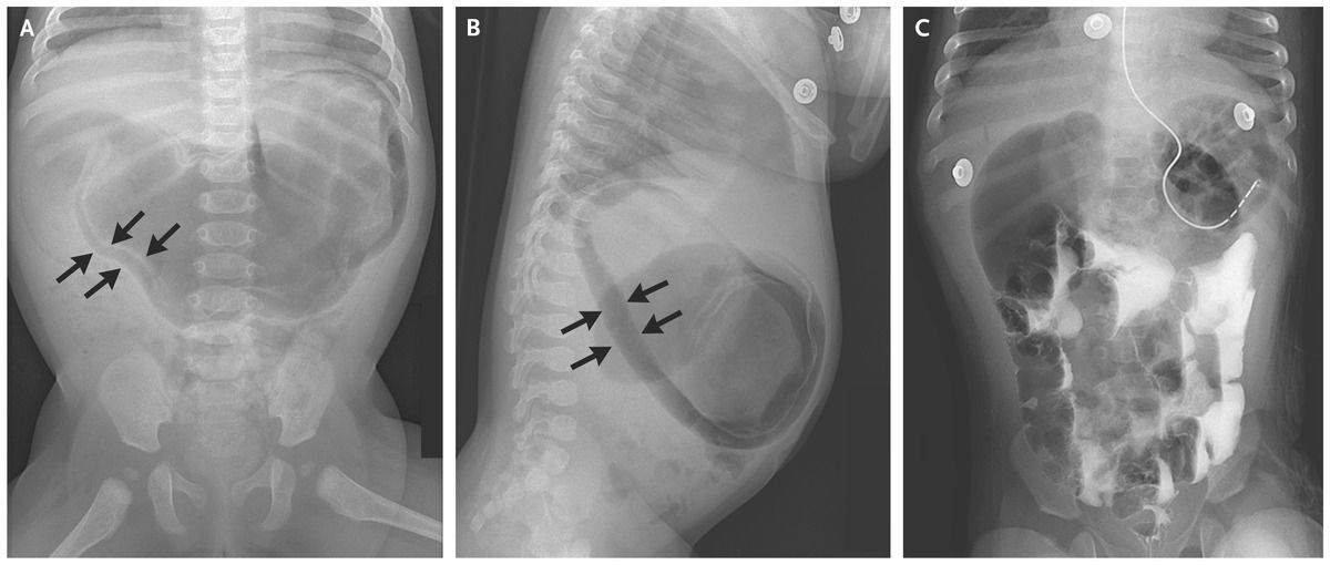
Laboratory investigations revealed a hypochloremic metabolic alkalosis, and radiographs showed gastric pneumatosis (Panels A and B, arrows).
A nasogastric tube was placed, and intravenous administration of fluids was started. Radiography of the upper gastrointestinal tract with contrast revealed contrast medium passing into the small intestine; follow-up images showed resolution of pneumatosis and a “double bubble,” which suggested duodenal obstruction (Panel C).
Duodenal obstruction caused by a duodenal web was identified intraoperatively, and a duodenoduodenostomy was performed, which resulted in resolution of symptoms.
What is Gastric pneumatosis ?
Gastric pneumatosis is rare and probably results from mucosal disruption due to ischemia or infection that allows gas to infiltrate into the wall of the stomach.
In newborns, gastric pneumatosis is associated with necrotizing enterocolitis, but increased intragastric pressure from severe obstruction caused by duodenal blockage, pyloric stenosis, or a lactobezoar may also produce gastric pneumatosis.


