This post is an answer to the ECG Case 222
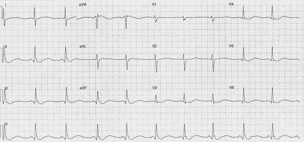
- Rate: 60 bpm
- Rhythm: Sinus arrhythmia
- Axis: Normal
- Intervals:
- PR – Normal (~150ms)
- QRS – Prolonged (120ms)
- Apparent QT – 680ms (QTc Bazette ~ 670 ms)
- Segments:
- ST Elevation lead aVR (1mm)
- ST Depression in leads II, III, aVF, V3-6
- Additional:
- Prominent U waves
- T-U Fusion
- Best visualised in leads II, III, aVF, V3-6
- Initial T wave is inverted and merges with large U wave
- Results in apparent QT prolongation due to fusion
- Best considered QU prolongation
Interpretation
Features consistent with hypokalaemia +/- hypomagnesaemia. The patient’s potassium was 2.2 mmol/L and magnesium 0.9 mmol/L.
READ MORE: Hypokalemia ECG Changes [With Examples]
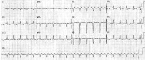
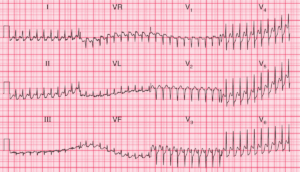
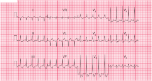
SIMILAR CASES: