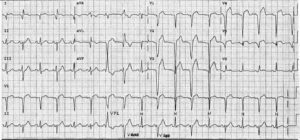
ECG Interpretation
- Sinus rhythm, rate 48/min
- Normal axis
- QRS complex duration normal, but the R wave height in lead V5 is 30 mm, and the S wave depth in lead V2 is 25 mm
- Inverted T waves in leads I, VL, V5–V6 (strain pattern)

Clinical Interpretation
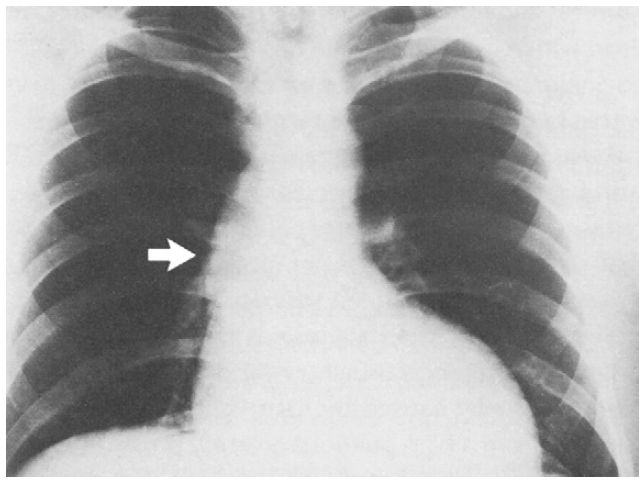
This is the classic ECG appearance of left ventricular hypertrophy. The chest X-ray showed an enlarged left ventricle with ‘post-stenotic’ dilatation of the ascending aorta (arrowed).
What to do next?
The combination of dizziness on exercise, a systolic murmur, and evidence of left ventricular hypertrophy suggests significant aortic stenosis.
The next step is an echocardiogram: in this patient it showed a gradient across the aortic valve of 140 mmHg, indicating severe stenosis. He needed an urgent aortic valve replacement.
- READ MORE about:
- SIMILAR CASES: