This post is an answer to the ECG Case 277
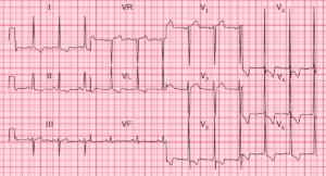
- Rate: 75 bpm
- Rhythm: Regular sinus rhythm
- Axis: Normal
- Intervals:
- PR – Short (100 ms)
- QRS – Prolonged (120 ms)
- QT – 380 ms (QTc Bazette 425 ms)
- Segments:
- ST Elevation in leads aVR, V1-3
- ST Depression in leads I, II, III, aVF, V5-6
- Additional:
- Delta Waves in leads I, II, III, aVF, V5-6
- Voltage criteria for LVH
- S wave in V1 + R wave in V5 > 35mm
- NOTE in the setting of pre-excitation this reflects the pre-excitated depolarisation rather than morphological LVH
Interpretation
- Wolff-Parkinson-White (WPW)
- PR Shortening
- QRS Prolongation
- Delta Waves
- Anteroseptal pathway (Arruda Algorithm)
- ST changes and apparent / ‘pseudo’ LVH due to pre-excitation
What happened next ?
The patient had a normal chest x-ray and a diagnosis of chest wall pain was made. There was no prior history of syncope or palpitations and she was referred for paediatric cardiology follow-up.
SIMILAR CASES: