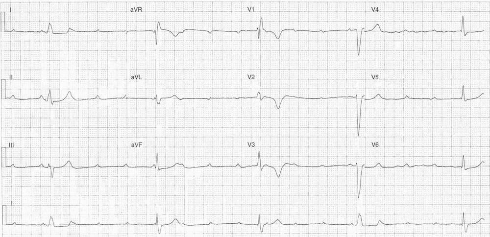
This post is an answer to the ECG Case 311
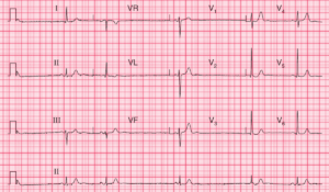
- Mean ventricular rate ~48 bpm
- Mean atrial Rate ~84bpm
- 2:1 AV block
- LBBB Morphology
- Sgarbossa negative
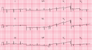
Another ECG recorded later
- Mean ventricular rate 30 bpm
- Mean atrial rate 102 bpm
- Complete heart block
- AV Dissoication
- Atrial rate > ventricular rate
- Complexes #1 & #4
- LBBB Morphology – same as ECG above
- Complexes #2, #3, & #5
- RBBB Morphology
- Deep T wave inversion leads V1-3
- ? Cardiac T wave memory
Pacemaker Insertion Indication
This patient has a clear indication for PPM insertion given the AV block seen on the ECGs. However even in the absence of AV block the second ECG also has another indication for PPM insertion. The second ECG also shows bilateral bundle branch block, evidence of both RBBB and LBBB, this is clear evidence of disease in all 3 fascicles.
What happened
The patient was admitted under the cardiology team and placed on an isoprenaline infusion. He underwent subsequent single chamber pacemaker insertion and was discharged following a brief in-patient stay.
READ ALSO: Conduction Blocks at the AV Node (AV Blocks) [With Examples]
SIMILAR CASES: