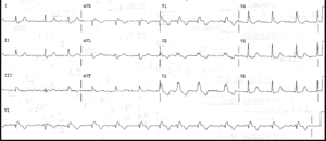
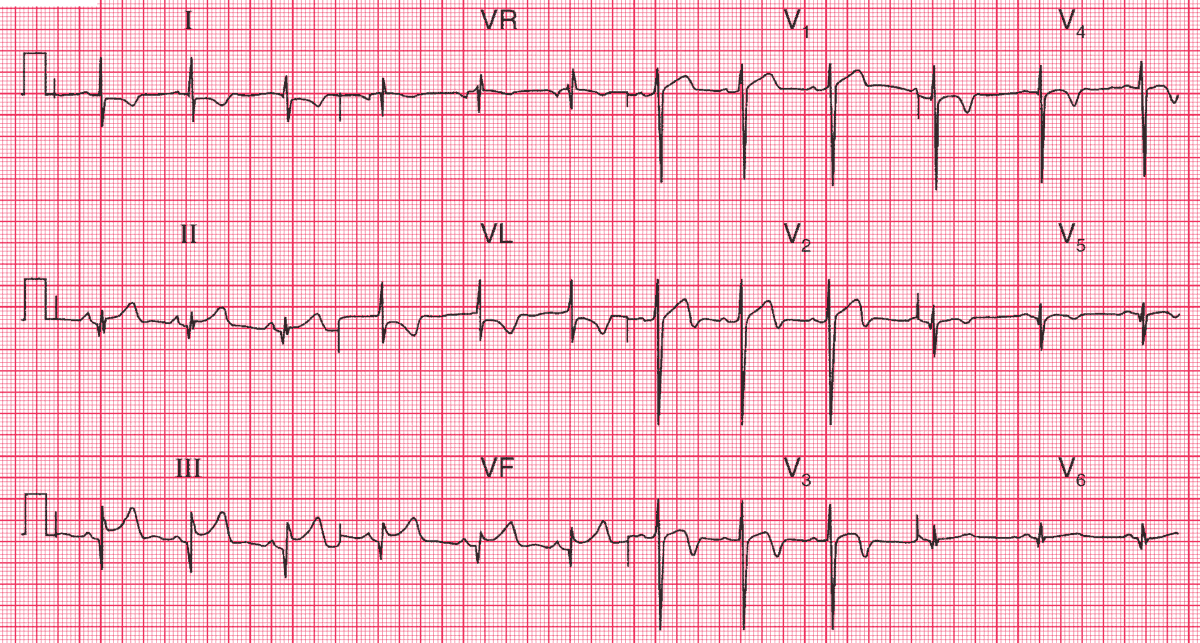
ECG Interpretation
- Sinus rhythm, average rate 70/min
- Normal axis
- Q waves in leads II, III, aVF (Possible old MI)
- Normal QRS complexes elsewhere
- Raised ST segments in leads II, III, aVF
- Reciprical ST segment depression in leads I and aVL
- Biphasic (up-down )T waves in leads V2–V3
- Inverted T waves in leads V4–V5
Clinical Interpretation
The inferior Q waves could suggest an old infarction. The raised ST segments in leads II, III and aVF would be compatible with an acute infarction. The anterior changes suggest anterior MI with possible reperfusion because of the biphasic (up-down ) T waves in leads V2–V3 and the inverted T waves in leads V4–V5.
What to do ?
There is enough evidence here from leads II, III and aVF to justify percutaneous coronary intervention (PCI) or thrombolysis – which should, of course, be combined with pain relief and aspirin.
- READ MORE:
- Similar Cases: