This post is an answer to the Case – 86-year-old Man With Dyspnea and Fatigue
Findings
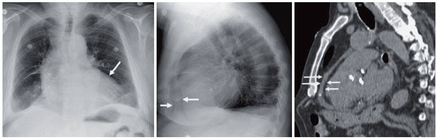
- Initial radiograph shows global enlargement of the cardiac silhouette without pulmonary vascular redistribution or pulmonary edema. On the frontal view, the cardiac silhouette is less dense along the left heart border (differential density sign) (arrow on frontal view radiograph).
- On the lateral view, two radiolucent arcs can be seen near the anterior chest wall (arrows). These represent the epicardial and pericardial fat separated by pericardial fluid. This finding has been referred to as the fat pad sign, sandwich sign, or Oreo cookie sign.
- CT reconstruction (asterisk = pericardial fluid; anterior arrows = pericardial fat; posterior arrows = epicardial fat) shows how the fat pads can create this sign.
Differential Diagnosis
Dilated cardiomyopathy or pericardial effusion can explain an enlarged cardiac silhouette without pulmonary edema or vascular redistribution. The presence of the sandwich sign confirms the latter.
Teaching Points
- Sonography is used for the initial detection and characterization of pericardial effusions.
- Although the water bottle appearance of the heart is well published, the ability to detect this configuration prospectively is somewhat limited.
- When global enlargement of the cardiac silhouette is encountered, comparison should be made to prior radiographs. Sudden changes in cardiac size should prompt one to consider pericardial effusion over cardiomyopathy.
- The differential density sign refers to decreased attenuation of the cardiac periphery from a pericardial effusion. As no heart is in this portion, the pericardial fluid attenuates the x-ray beam less than the central portion.
- On the lateral radiograph, the fat pad sign may be seen. Normally the pericardial stripe should be only 2 to 4 mm.
- Pericardial effusions may be secondary to collagen vascular diseases (especially lupus), myxedema, uremia, radiation, and infection (viral or mycobacterial). Occasionally, they may be malignant or sanguineous (hemopericardium).
Management
- The initial finding of global enlargement of the cardiac silhouette on a chest radiograph should prompt an echocardiogram.
- Management is based on the size of the effusion and its effect on the heart.
SIMILAR CASE: Uremia and Shortness of Breath
Further Reading
Woodring JH. The lateral chest radiograph in the detection of pericardial effusion: a reevaluation. J Ky Med Assoc. 1998 Jun;96(6):218-24. PMID: 9642917.