This post is an answer to the Case – 55-year-old Man With Chest Discomfort
Findings
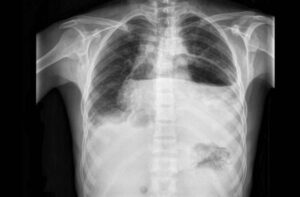
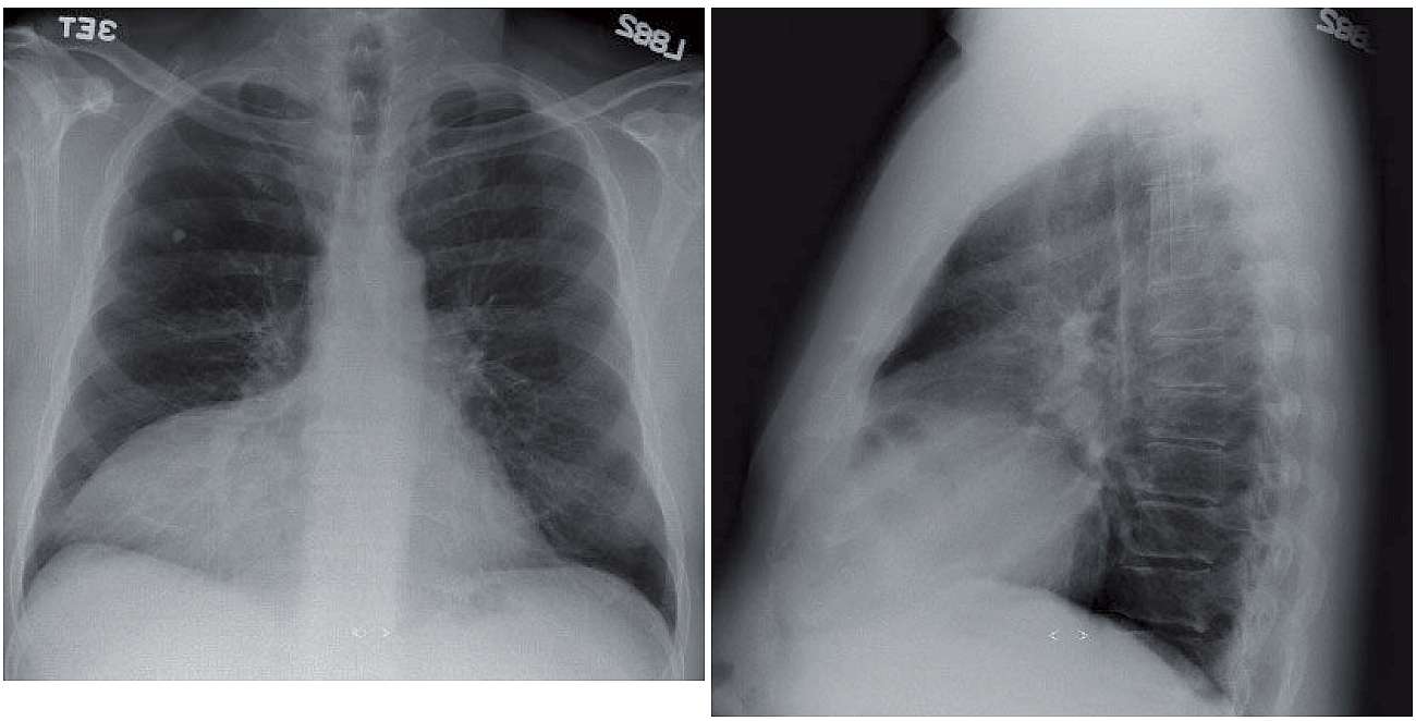
- Right cardiophrenic angle soft tissue mass is seen on the frontal radiograph, obscuring the right heart border. Right interlobar and lower lobe pulmonary arteries can still be seen (hilum overlay sign, suggesting anterior mediastinal process).
- On the lateral radiograph, the soft tissue mass is located anteriorly and contains a loop of partially air-filled bowel.
Differential Diagnosis
Foramen of Morgagni hernia, pericardial cyst, abundant pericardial fat, lymphadenopathy/lymphoma, or other anterior mediastinal mass (thymoma, germ cell tumor) should be considered when a right cardiophrenic angle mass is encountered. Identification of the bowel loop makes the foramen of Morgagni the most likely diagnosis.
Teaching Points
- Resulting from a defect in the attachment of diaphragmatic muscle fibers to the costal margin and central tendon of the diaphragm, foramen of Morgagni hernias are relatively rare (<3 % diaphragmatic hernias).
- They are more commonly right-sided, and usually contain herniated omentum or portions of the transverse colon.
Management
- CT can be used to confirm the diagnosis and exclude other differential entities when the diagnosis is not clear on the radiograph. Herniation of omental fat into the retrosternal space is noted at CT and can be differentiated from abundant pericardial fat by omental vessels coursing through the fat or herniated abdominal viscera.
- Surgical consultation is recommended, as there is a risk of abdominal organ incarceration if left unrepaired. A transabdominal surgical approach is now preferred.
SIMILAR CASE: An 8-year-old Girl with Light Cough when Laying Down
Further Reading
Minneci PC, Deans KJ, Kim P, Mathisen DJ. Foramen of Morgagni hernia: changes in diagnosis and treatment. Ann Thorac Surg. 2004 Jun;77(6):1956-9. doi: 10.1016/j.athoracsur.2003.12.028. PMID: 15172245.