ECG Interpretation
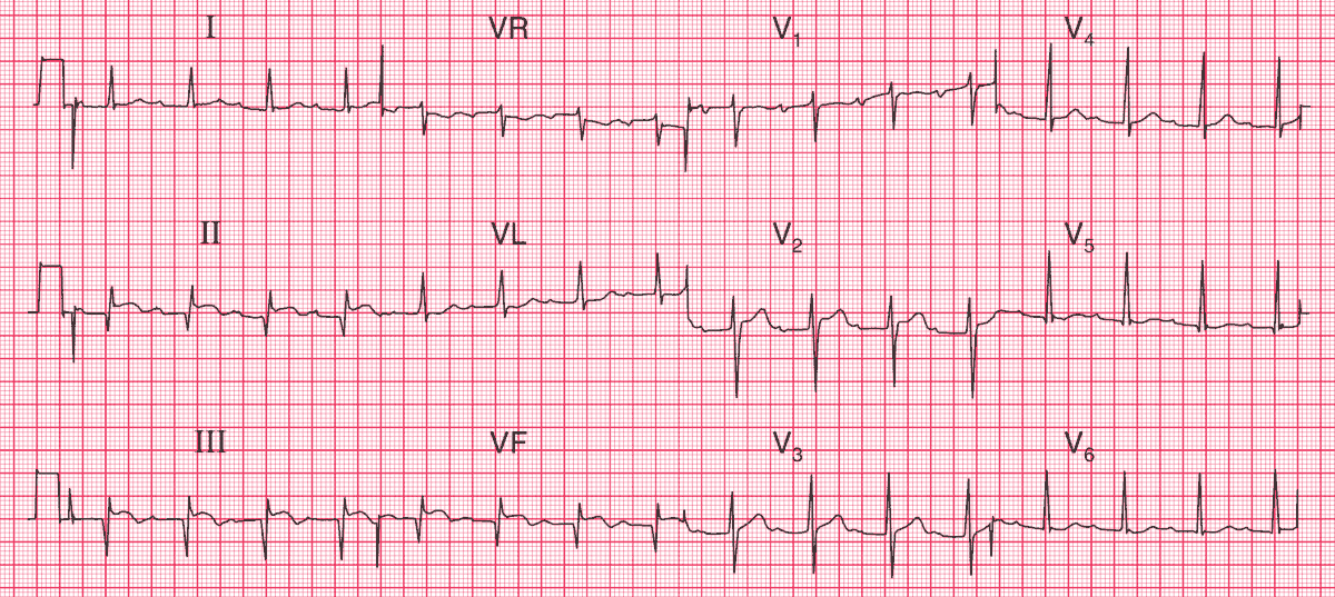
- Sinus rhythm, rate 88/min
- PR interval 320 ms – first degree AV block
- Q waves in leads II, III, VF
- Raised ST segments in leads II, III, VF
- Inverted T waves in leads III, VF
Clinical Interpretation
This ECG shows an acute inferior myocardial infarction, which often causes first degree block. The Q waves and raised ST segments are consistent with the story of 6 h of chest pain, and the first degree block is not important.
What to do next?
Chest pain radiating through to the back has to raise the possibility of aortic dissection, which can occlude the opening of the coronary arteries and so cause a myocardial infarction. However, this is relatively rare compared with back pain associated with myocardial infarction, which is common.
In this case, the chest X-ray showed that blood has leaked into the left pleural cavity from a dissection of the aorta. Thrombolysis for the myocardial infarction is obviously contraindicated, and the patient needs immediate investigation by CT or MR scanning to see if surgical repair of the dissection is possible.
- READ MORE:


