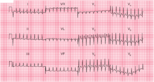
ECG Interpretation
- Atrial fibrillation, with one ventricular extrasystole
- Normal axis
- Normal QRS complexes
- Horizontal ST segment depression of 7 mm in lead V2
- Downward-sloping ST segment depression in leads V3–V6, maximal in leads V3 and V4
- Tall R waves in precordial leads (posterior Q waves)
- Inverted T waves in leads I, VL, V6; indeterminate T waves elsewhere
Clinical interpretation
The anterior horizontal ST segment depression maximal in leads V3 and V4 and the tall R waves indicates posterioir MI. The ventricular rate is not too fast, and although the heart rate may be contributing to the ischaemia it seems unlikely that it is the main problem.
What to do ?
The patient should be treated for an acute coronary syndrome.If the pain does not settle, early angiography with a view to revascularization by a coronary artery bypass graft (CABG) or percutaneous coronary intervention (PCI) will have to be considered.