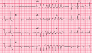
ECG Interpretation
- Sinus rhythm, rate 81/min
- Markedly peaked P waves (best seen in leads II, V1)
- Normal axis
- Dominant R wave in lead V1
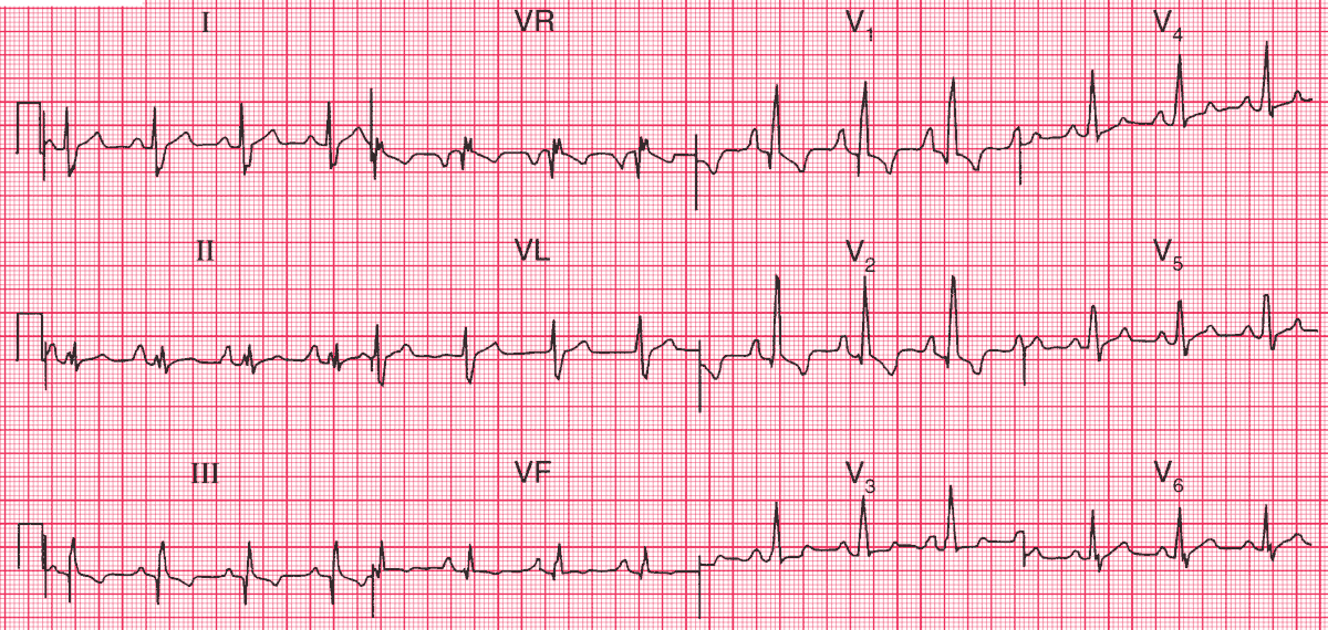
Clinical Interpretation
The ECG shows right atrial and right ventricular hypertrophy.
What to do ?
Right atrial hypertrophy is seen with pulmonary hypertension of any cause, tricuspid stenosis, and Ebstein’s anomaly. Right ventricular hypertrophy is seen with pulmonary stenosis and pulmonary hypertension. These conditions can all be diagnosed by echocardiography. This patient had pulmonary valve stenosis.