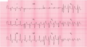
ECG Interpretation
- Sinus rhythm, rate 140/min
- One ventricular extrasystole
- Peaked P waves best seen in leads II, III, VF (P pulmonale) – a sign of Right Atrial Enlargement (RAE)
- Inverted P waves in lead aVL
- Right axis deviation
- Poor R wave progression in precordial leads (delayed R/S transition)
- Deep S wave in lead V6
- Normal ST segments and T waves
Clinical Interpretation
The sinus tachycardia suggests a major problem. The peaked P waves indicate right atrial hypertrophy. The right axis deviation and dominant R wave in lead V1 suggest right ventricular hypertrophy. The deep S wave in lead V6, with no ‘left ventricular’ complexes in the chest leads, indicates ‘clockwise rotation’ of the heart, with the right ventricle occupying the precordium. These changes suggest lung disease.
What to do?
Since the ECG is entirely ‘right-sided’, one can assume that the problem is due to chronic lung disease or recurrent pulmonary embolism. The story sounds more in keeping with a lung problem. The raised jugular venous pressure is presumably due to cor pulmonale. The sinus tachycardia is worrying, and may suggest respiratory failure.
- READ MORE: ECG Changes in Chronic Obstructive Pulmonary Disease (COPD)
- More Similar Cases: