The ECG shows a regular rhythm at a rate of 130 bpm. The QRS complex has a normal duration (0.10 sec), although in lead V1 it appears to be longer 0.14 sec, and has a morphology suggestive of right bundle branch block (RSR′ morphology in lead V1). However, the S waves in leads I and V5-V6 are not as wide as the R′ waveform in lead V1 (←).
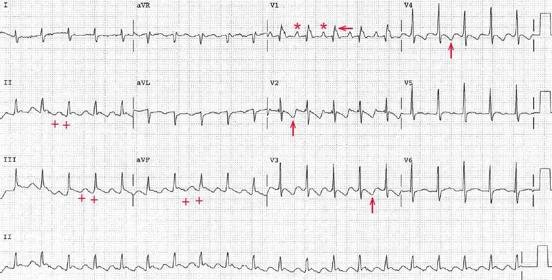
The axis is normal (+90°), with a biphasic QRS complex in lead I and a positive QRS complex in lead aVF. Although no distinct P waves are seen in leads II, III, and aVF, the baseline appears to have undulations (saw-tooth–like) that are suggestive of flutter waves (+).
In lead V1 a distinct atrial waveform can be seen before each QRS complex (*). The terminal portion of the R′ waveform in lead V1 has obvious notching (↓), suggesting another atrial waveform. Indeed, when the intervals are measured they are all regular. The QT/QTc intervals are normal (280/410 msec).