This article is an answer to the ECG Case 184
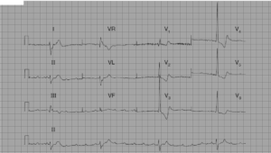
The ECG shows there is a regular rhythm at a rate of 100 bpm. There is variability of the QRS complex morphology and width; there is repeating pattern of group beating consisting of three wider QRS complexes (↑) with a duration of 0.18 sec and one narrower QRS complex (↓) (0.14 sec) that also has a different amplitude.

There is a tall R wave in lead V1 (→) and the R wave is broader than the S wave, a feature associated with a ventricular complex. In addition, the QRS complexes do not have either a right or left bundle branch block morphology. Therefore, this is an accelerated idioventricular rhythm, also called slow ventricular tachycardia.
There are no P wave seen before any of the wider QRS complexes, but there is a P wave (+) after the third wider QRS complex and before the narrower QRS complex (↓). The P wave is negative in leads I, II, aVF, and V4–V6. Therefore, it is not originating from the sinus node.
Negative P waves are either retrograde due to VA conduction or a low atrial focus. As this is a ventricular rhythm, this is most likely a retrograde P wave due to VA conduction. Although there are no other obvious P waves seen associated with the wider QRS complexes, noted are changes in the T waves in the lead II rhythm strip that usually indicate superimposed P waves.
The first wider QRS complex in the lead II rhythm strip has a deep and narrow negative waveform (^), and the second wider QRS complex has a negative notching on the upstroke of the T wave (*). These P waves also appear to be negative. The P waves are best seen in lead V1. As they are seen with the wider QRS complexes, they are not premature atrial waves, but rather they are associated with the QRS complex. Hence this is a ventricular rhythm with retrograde P waves as a result of VA conduction.
However, it can be noted that there is a progressive lengthening of the RP interval (└┘) from 0.32 to 0.40 to 0.48 sec, ie, retrograde Wenckebach. This occurs in a repeating pattern. The retrograde P wave after the third wide QRS complex (+) results in the narrower QRS complex (↓) that is difference in morphology from the other three QRS complexes. Indeed, it has a more normal looking morphology in the precordial leads with normal R wave progression. It is in response to the retrograde P wave and is an echo beat.
READ MORE:
SIMILAR CASES: