This post is an answer to the ECG Case 217
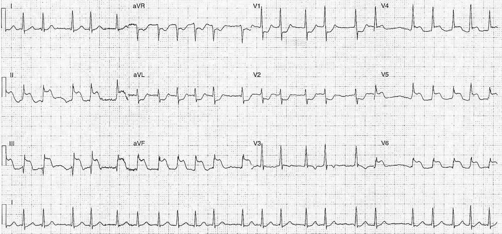
- Rate: Mean rate ~130bpm
- Rhythm:
- Irregular
- Nil p waves
- Axis: Normal
- Intervals:
- PR – Nil p waves
- QRS – Normal (80ms)
- QT – 280ms
- Segments:
- ST Elevation in leads II (4mm), III (5mm), aVF (4mm), V4 (2mm), V5 (3mm), V6 (2.5mm)
- ST Depression in leads aVL, aVR, V1-2 (note horizontal morphology)
- Additional:
- Prominent R wave V1-2
Interpretation
Infero-lateral-posterior OMI with Atrial Fibrillation
What happened next?
The ECG findings were immediately recognised and local PCI protocol was activated. The angiogram showed:
- LAD – 70% mid-stenosis
- PLA – 100% ostial occlusion –> Stented
- LV function preserved
Echocardiogram:
- Inferolateral LV akinesis
- Normal RV size and function
The patient’s recovery was complicated by acute stent thrombosis requiring re-stent. He was discharge following a 4 day in-patient stay.
READ MORE:


