This post is an answer to the ECG Case 286
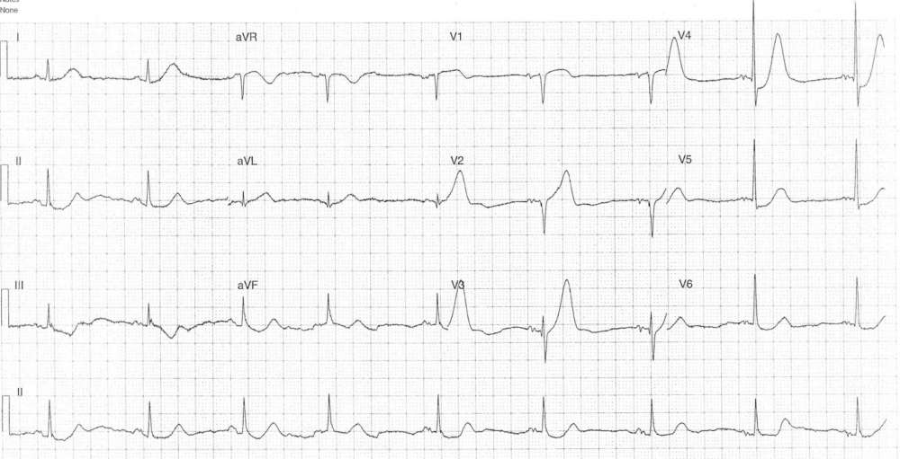
- Rate: 54 bpm
- Rhythm: regular sinus rhythm
- Axis: Normal
- Intervals:
- PR – Normal (~180ms)
- QRS – Normal (100ms)
- QT – 440ms
- Segments:
- ST Elevation in leads aVR, V1 (1-1.5mm), V2 (<1mm)
- ST Depression in leads I, II, V4-6
- Additional:
- Marked hyperacute T waves in leads V2-4
- Some ST segment analysis is difficult due to baseline artifact
- T wave inversion in lead III
Interpretation
- De Winter’s Pattern
- Hyperacute T waves with associated ST depression in leads V2-6
- Possible ST elevation aVR
What happened next ?
The patient went for urgent angiography which showed:
- LM: Normal
- LAD: Proximal 80% Mid 70% stenosis
- LCx: moderate diffuse disease
- RCA (Dominant): Mild 50%, Distal 70% PLV 80%
- LV: Anterior hypokinesia with mild-mod LV dysfunction
The LAD lesion under DES PCI and the patient returned to hospital 2 months later for an elective staged PCI to the RCA.
READ MORE: ECG Interpretation – All you need to know


