This post is an answer to the ECG Case 338
- Rate: 48 bpm
- Rhythm: Regular, sinus rhythm
- Axis: Normal
- Intervals:
- PR – Normal (~160ms)
- QRS – Normal (100ms)
- QT – 480ms (QTc Bazette 430 ms)
- Note the ECG machine calculated the QT 500ms / QTc 480ms
- Likely due to low T wave voltage plus T-U fusion
- Additional:
- Subtle ST depression in leads II, aVF
- U wave visible in leads V2-3
- T waves globally flat
- Relatively long ST segment
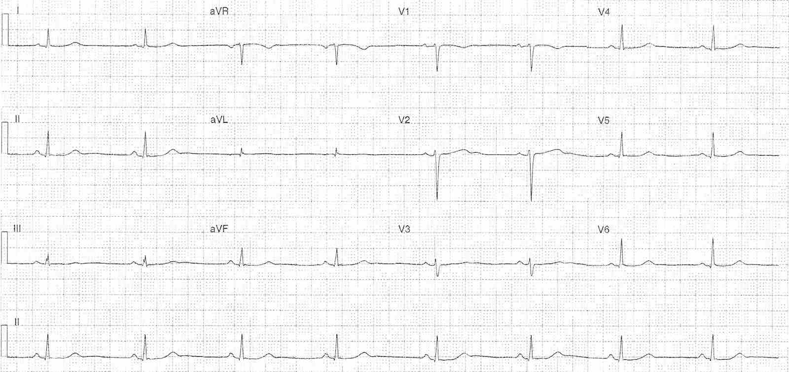
Changes consistent with electrolyte abnormality – differentials:
- Hypokalaemia – U waves, T-U fusion with QT prolongation, ST depression, low voltage T waves
- Hypomagnesaemia – associated with hypokalaemia
- Hypocalcaemia – causes long QT due to ST segment lengthening
What happened next ?
The patient’s potassium (K) was 2.7 mmol/L with a low-normal magnesium and low-normal calcium. Following IV electrolyte replacement the patients ECG changes resolved and her symptoms of weakness also resolved.
READ ALSO:
SIMILAR CASES: