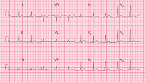
The ECG shows a regular rhythm at a rate of 130 bpm. P waves are not obvious in most leads. However, a positive P wave can be seen before each QRS complex in leads V4-V6 (*).
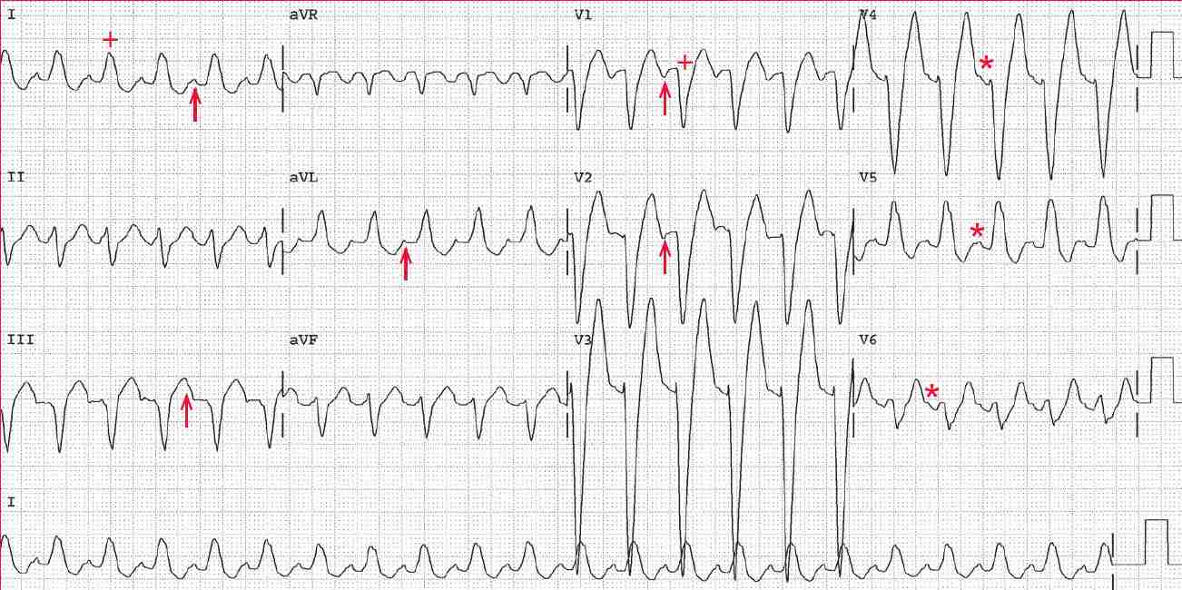
The PR interval is stable (0.14 sec). Using this PR interval, it can be seen that the negative deflection noted in leads V1-V2 (↑) as well as the positive deflection (↑) in leads I, aVL, and III are the P waves. This is, therefore, sinus tachycardia.
The QRS complex duration is prolonged (0.16 sec), and there is a pattern characteristic of left bundle branch block (LBBB; broad R wave in leads I and V5 and a QS complex in lead V1 [+]).
The axis in the frontal plane is leftward (positive QRS complex in lead I and negative QRS complex in leads II and aVF). The QT/QTc intervals are prolonged (320/470 msec) but are normal when corrected for the prolonged QRS complex duration (240/360 msec).